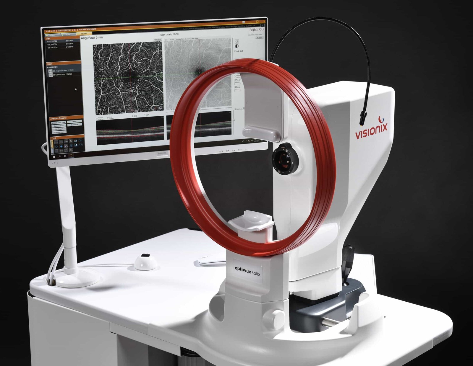
The leading Optovue Solix alternatives include ZEISS CIRRUS 6000 with AngioPlex, Topcon DRI OCT Triton with OCT-A, and Heidelberg SPECTRALIS with its OCT Angiography Module. Each offers different imaging technology, clinical analysis, and pricing options. The best choice depends on your patients, practice needs, and equipment budget.
Your next OCT could change how your clinic sees patients. Want more insight per scan? Compare Solix, ZEISS, Topcon, and Heidelberg, then choose the best-fit system for your practice today.
First, let’s look at a few questions many may ask.
What Is the Difference Between OCT and OCT-A?
Optical coherence tomography (OCT) shows the layers and structure of eye tissue. OCT angiography (OCT-A) adds maps of blood flow without injecting dye. Both help clinicians assess:
- Retinal disease
- Glaucoma
- Other conditions
However, each technology answers different clinical questions.
Structural OCT measures:
- Tissue thickness
- Shape
- Changes over time
OCT-A displays flow signals within retinal and choroidal vessels. It can help assess microvascular changes but does not replace fluorescein angiography when dye studies are needed.
Image artifacts and segmentation errors still require careful review.
A useful purchase begins with your exam mix. For example:
- A busy glaucoma service may value repeatable nerve fiber layer reports.
- A retina clinic may prioritize vascular maps and deeper choroidal imaging.
- A cornea-focused clinic may need strong anterior segment tools.
Understanding your most common clinical needs helps you select equipment that delivers lasting value.
How Much Does an OCT-A Machine Cost?
An OCT-A quote reflects far more than the scanner. Software access, lenses, training, computer hardware, service coverage, and shipping can change the total. An attractive base price is only useful when the included features meet your needs.
For a real-world snapshot, Ferris Optical listed the following equipment prices currently:
- Optovue Solix OCT / OCT-A: $34,999
- Topcon DRI OCT Triton Plus: $17,999
- Heidelberg SPECTRALIS OCT2 Module: $19,900
These are listed asking prices, not matched quotes for identical OCT-A configurations. Stock and pricing may change.
Be sure to ask whether the quoted system includes:
- Angiography activation
- The required workstation
- Current software
The Heidelberg OCT2 listing describes a module, so buyers must verify the host platform and OCT-A module. A low sticker price should not hide necessary upgrades.
Optovue Solix: The Starting Point
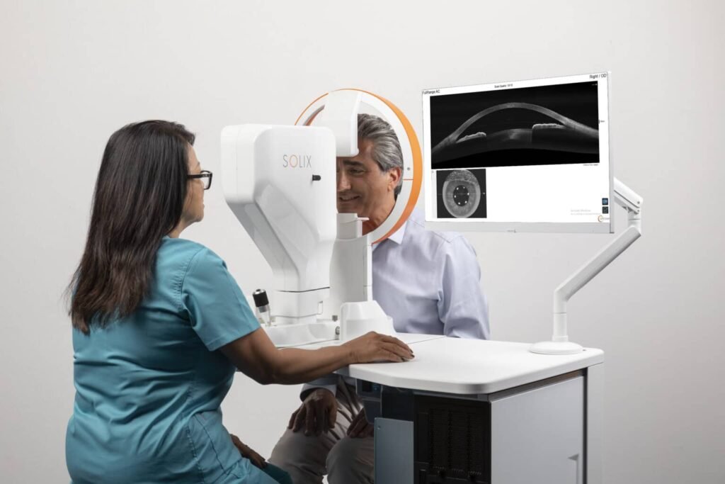
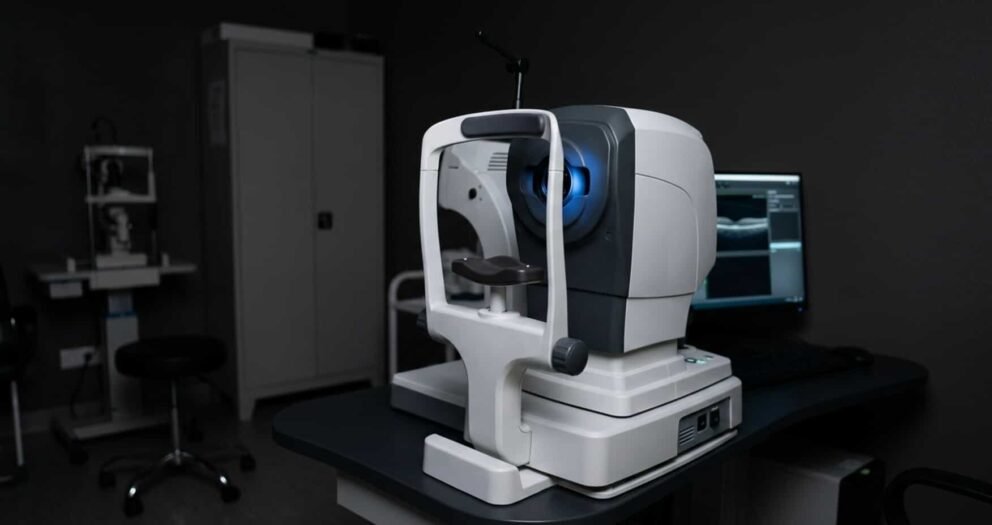
Optovue Solix uses spectral-domain OCT (SD-OCT) and the FullRange platform. It has 120,000 A-scans per second and supports retinal scans up to 16 mm wide.
FullRange depth reaches 6.25 mm in supported modes. The system covers:
- Retinal structure
- OCT-A
- Glaucoma analysis
- Anterior segment imaging
AngioVue provides vascular imaging and analysis. Available protocols include 3 × 3, 6.4 × 6.4, 9 × 9, and 12 × 12 mm retinal OCT-A scans.
Montage views can extend coverage.
Corneal mapping, pachymetry, epithelial analysis, and chamber imaging add value for practices serving both medical and primary eye care patients.
Solix configurations are not identical. Essential and more complete AngioVue packages have different analysis tools.
Be sure to confirm the installed version before treating every advertised feature as standard. The configuration matters as much as the model name.
Optovue Solix vs ZEISS CIRRUS 6000
The Optovue Solix vs ZEISS CIRRUS 6000 decision often turns on reporting needs and existing clinical systems. Both use spectral-domain OCT. CIRRUS 6000 records 100,000 A-scans per second and offers AngioPlex OCT angiography.
Solix is rated at 120,000 A-scans per second, but scan rate alone does not predict image quality or exam time.
Retinal and Glaucoma Analysis
CIRRUS offers macular and optic disc cubes, ganglion cell analysis, retinal nerve fiber layer measurements, and Guided Progression Analysis. Its reference databases and established report formats may suit clinics that track glaucoma across many visits.
AngioPlex adds retinal perfusion and optic nerve head vascular views with a license.
For retina, CIRRUS supports 3 × 3 through 12 × 12 mm OCT-A scans, plus larger montage views. FastTrac eye tracking helps control motion during capture.
Solix counters with:
- AngioVue analysis
- Integrated wellness protocols
- Broad anterior segment imaging
Be sure to review sample reports from both before choosing.
Anterior Segment and Workflow
CIRRUS can support corneal thickness, angle, and anterior chamber imaging with the proper lens and license. Solix places stronger emphasis on corneal layer maps and FullRange anterior imaging. Practices already using ZEISS review tools may value continuity of records and staff familiarity.
Confirm data migration options before replacing an older system.
Optovue Solix vs Topcon Triton
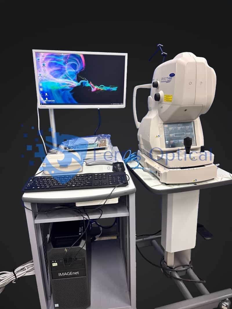
The main difference between Optovue Solix and Topcon Triton is the light source. Triton uses swept-source OCT (SS-OCT) at 1,050 nm. Solix uses spectral-domain OCT.
The longer wavelength can help reveal deeper structures such as the choroid. It can also support imaging through some media opacities.
Deep Retinal Imaging and Multimodal Views
Topcon lists 100,000 A-scans per second and a 12 × 9 mm wide structural scan. Triton combines OCT with true-color fundus photography and digital red-free images.
Triton Plus can also add fluorescein angiography and fundus autofluorescence. Those dye and autofluorescence features are separate from OCT-A. With SS-OCT Angio and OCTARA processing, suitable Triton configurations show vascular flow at different retinal depths.
Retina specialists managing macular disease or choroidal findings may find the deeper structural view useful. Solix remains appealing when corneal mapping and glaucoma screening share the same exam room.
Glaucoma, Anterior Imaging, and Patient Flow
Topcon offers optic disc scans and imaging for glaucoma follow-up. Optional anterior segment imaging broadens its reach. An invisible scan beam and tracking tools can help patients hold fixation.
Compare the exact scan protocol, OCT-A access, and analysis package on the used unit. Do not assume a Triton Plus includes every optional module.
Optovue Solix vs Heidelberg SPECTRALIS
Buyers researching Optovue Solix vs Heidelberg should first examine the installed modules. SPECTRALIS is a flexible imaging platform rather than one fixed package.
A system with OCT2 hardware may not include OCT angiography, glaucoma analytics, or anterior imaging unless those features are present and enabled.

Repeatable Imaging for Long-Term Follow-Up
SPECTRALIS uses spectral-domain OCT with TruTrack active eye tracking and AutoRescan tools. Its glaucoma module can align scans using anatomical landmarks.
Consistent scan placement helps clinicians compare the same tissue over time. Retina and glaucoma specialists may value that follow-up method. The OCT Angiography Module adds vessel imaging and segmentation tools.
Newer SHIFT-capable configurations offer different OCT-A scan speeds, including 85, 125, and 250 kHz modes. However, the listed SPECTRALIS OCT2 module is described at up to 85 kHz.
Never assign newer module speeds to an older unit without proof of its installed configuration.
Multimodal Growth and Anterior Options
SPECTRALIS can combine OCT with other retinal imaging modules. Its Anterior Segment Module provides detailed cornea and anterior chamber angle views.
Solix may offer a simpler all-in-one path for clinics that need frequent corneal measurements. Heidelberg can suit teams building a modular retinal imaging program over time.
OCT / OCT-A Systems Comparison: Which Features Matter Most?
A practical OCT / OCT-A systems comparison should focus on clinical questions, not product slogans. Scan speed, scan area, software tools, and patient positioning all affect daily use. Be aware that device labels never tell the entire story.
Match the Imaging Method to the Patient
For deep choroidal views, swept-source Triton has a clear technical distinction. For structural glaucoma trends, compare CIRRUS progression reports with SPECTRALIS repeat-scan alignment. Also, for corneal layers and mixed anterior-posterior care, review Solix FullRange tools.
Each choice offers useful strengths for a defined workflow.
Compare Output, Not Just the Screen
Ask vendors to show a macular cube, optic disc report, OCT-A slab, and follow-up trend from each device. Look for:
- Usable segmentation
- Easy review
- Clear printouts
Check whether images can move into your practice management or image archive system. A scanner that fits existing records may save staff time every day.
OCT Angiography Equipment Comparison: Check the Details
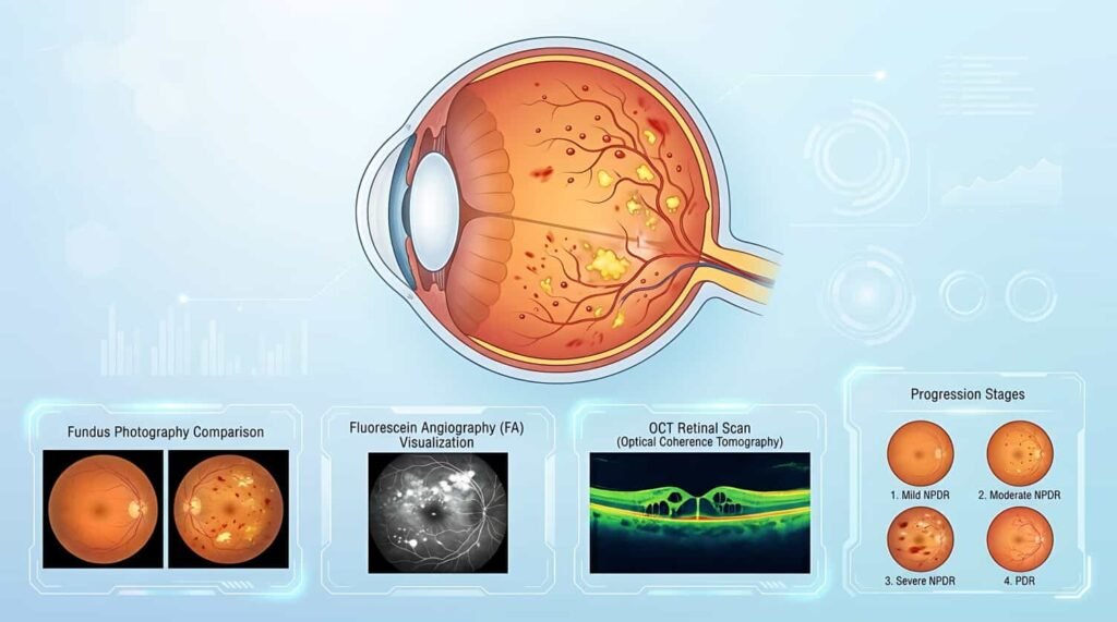
A serious OCT angiography equipment comparison must examine what the clinic can actually measure. OCT-A packages may differ in:
- Retinal scan size
- Vessel-density metrics
- Projection artifact removal
- Optic disc analysis
A device may produce attractive images without the quantitative report your protocol requires.
Request a live demonstration with the installed software. Test:
- Small-vessel detail
- Larger scan coverage
- Motion correction
- Manual segmentation review
- Export options
Ask how artifacts are marked. Compare scans from similar patients rather than comparing only brochure images.
Long-term trends need special care. OCT-A metrics often use each manufacturer’s algorithms and boundaries. Values from two different manufacturers may not be interchangeable.
Create a new baseline when switching platforms, and document the change in the patient record.
Best OCT Machines for Optometry Practices: Choose by Case Mix
The search for the Best OCT machines for optometry practices has no universal winner.
Start with the visits your clinic handles most often. Then select the imaging features your clinicians will use every week.
Consider the following practice needs:
- Mixed optometry and corneal care: Consider Solix for combined retinal, OCT-A, and anterior segment tools.
- Glaucoma-heavy care: Compare ZEISS CIRRUS 6000 reporting with Heidelberg SPECTRALIS follow-up alignment.
- Retina and choroid focus: Assess Topcon Triton swept-source depth and Heidelberg multimodal options.
- Growing specialty service: Consider available upgrades, service access, and room layout before purchase.
Allow technicians to test setup, fixation, image quality checks, and report review. Count rescans and exam minutes during a realistic demonstration.
Used OCT Equipment: Protect Your Budget and Your Workflow
Used OCT equipment can provide advanced imaging without the full cost of a new installation. Pre-owned OCT machines still require careful checks before purchase.
Request:
- The serial number
- Year
- Service history
- Software version
- A current demonstration
Ask for a written list of included items.
A complete workstation, keyboard, monitor, patient support, licenses, external lenses, and backup media may be important. Have the seller explain:
- Installation
- Calibration
- Shipping protection
- Service availability
- Warranty terms
Ferris Optical lists a contact address in Singapore and serves eye care buyers internationally. For a cross-border order, confirm:
- Freight terms
- Import duties
- Voltage
- Local service support
- Delivery timing
Product photos and a quoted price cannot replace a full equipment checklist.
Verify Software and Ownership Rights
OCT-A software and licensing deserve written confirmation.
Ask whether angiography, advanced glaucoma reports, review stations, and analysis modules are activated. Check if licenses transfer to a new owner and whether upgrades require manufacturer approval. Also, plan the full OCT / OCT-A machine cost across several years.
Add:
- Routine service
- Computer replacements
- Training
- Licenses
- Possible repair costs
Review the Optovue Solix price against other systems only after matching each included feature. An itemized quote makes the comparison fair.
Frequently Asked Questions
Can a Used OCT System Keep Existing Patient Records?
Sometimes, but the answer depends on the model, database, and software version.
Ask whether the scanner can import prior studies or access records through a compatible review station. Confirm DICOM support, backup formats, and user permissions before moving files.
Request a test transfer using de-identified records. Even if images migrate, analysis trends may not transfer between brands.
Keep the original studies available for review and note when a new measurement baseline begins. Before removing the previous system, verify that your team can still access historical scans for future patient evaluations.
How Can a Clinic Test an OCT-A System Before Buying?
Request a live demonstration with your usual exam protocols.
Include a macular cube, nerve fiber layer report, vascular scan, and one follow-up comparison. Watch how often the operator needs another scan. Review images on the reading workstation, not only the capture monitor.
Ask a retina or glaucoma specialist to assess segmentation and report usefulness.
What Should Be Included in an OCT Purchase Agreement?
The agreement should name the exact model, condition, serial number, accessories, software, and license status.
It should also define:
- Delivery
- Installation
- Testing
- Training
- Warranty coverage
- Who handles repairs
For international shipments, record freight terms and who is responsible for duties.
Ask how a failed acceptance test will be resolved. Written details protect your budget and help your team begin patient imaging with fewer surprises. Clear acceptance criteria also help ensure the delivered equipment matches the specifications and features agreed upon before purchase.
Find the Right Optovue Solix Alternatives: Shop Now
The right Optovue Solix alternatives support your clinical goals and equipment budget. Compare verified imaging features, active licenses, and ownership costs before deciding.
Ferris Optical helps eye care professionals worldwide equip their clinics with dependable new and pre-owned devices from leading ophthalmic manufacturers at competitive prices.
Explore the available OCT systems, request a clear equipment quote, and shop now at Ferris Optical to equip your practice.
 English
 Spanish
 German
 French