
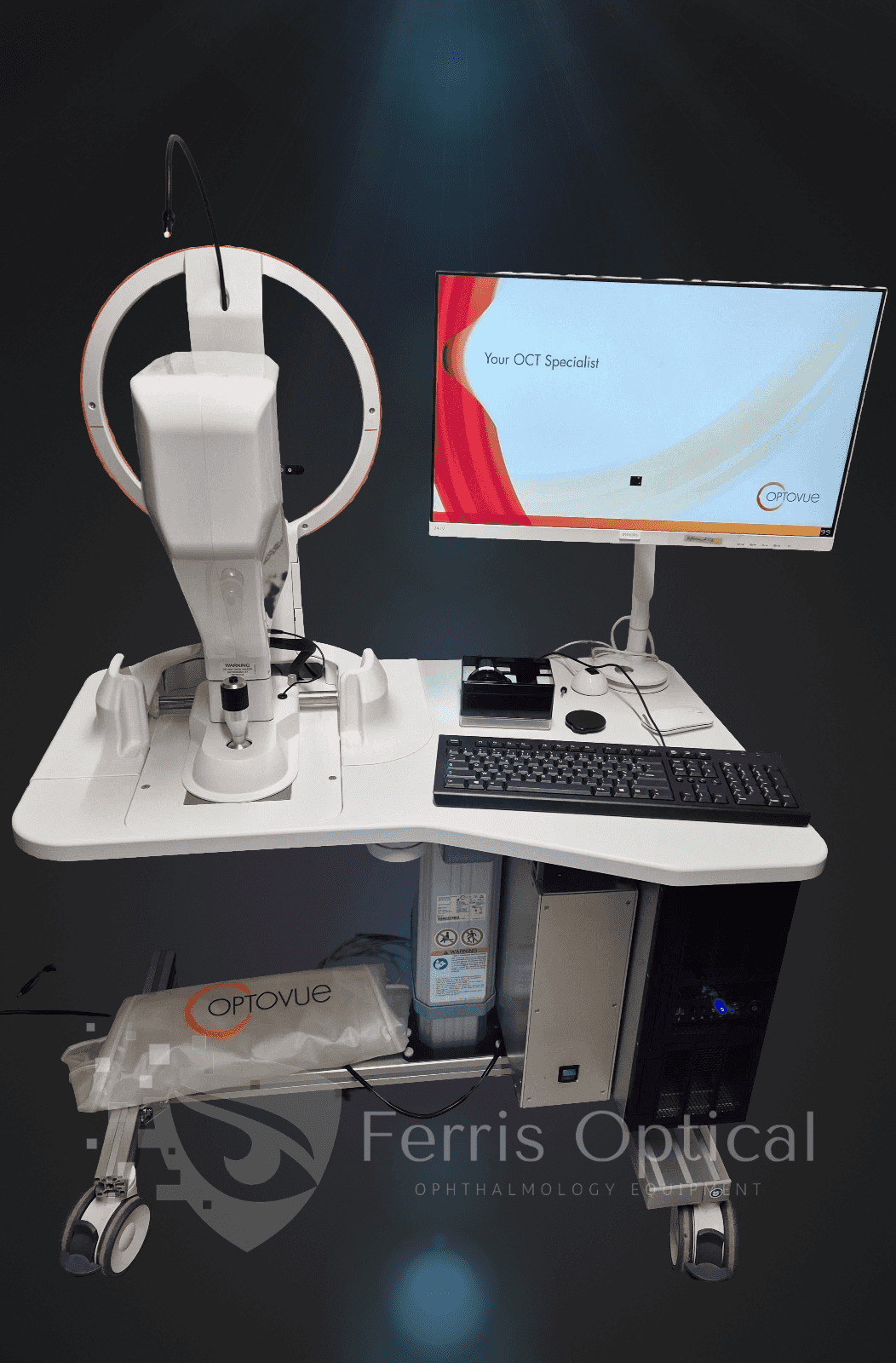
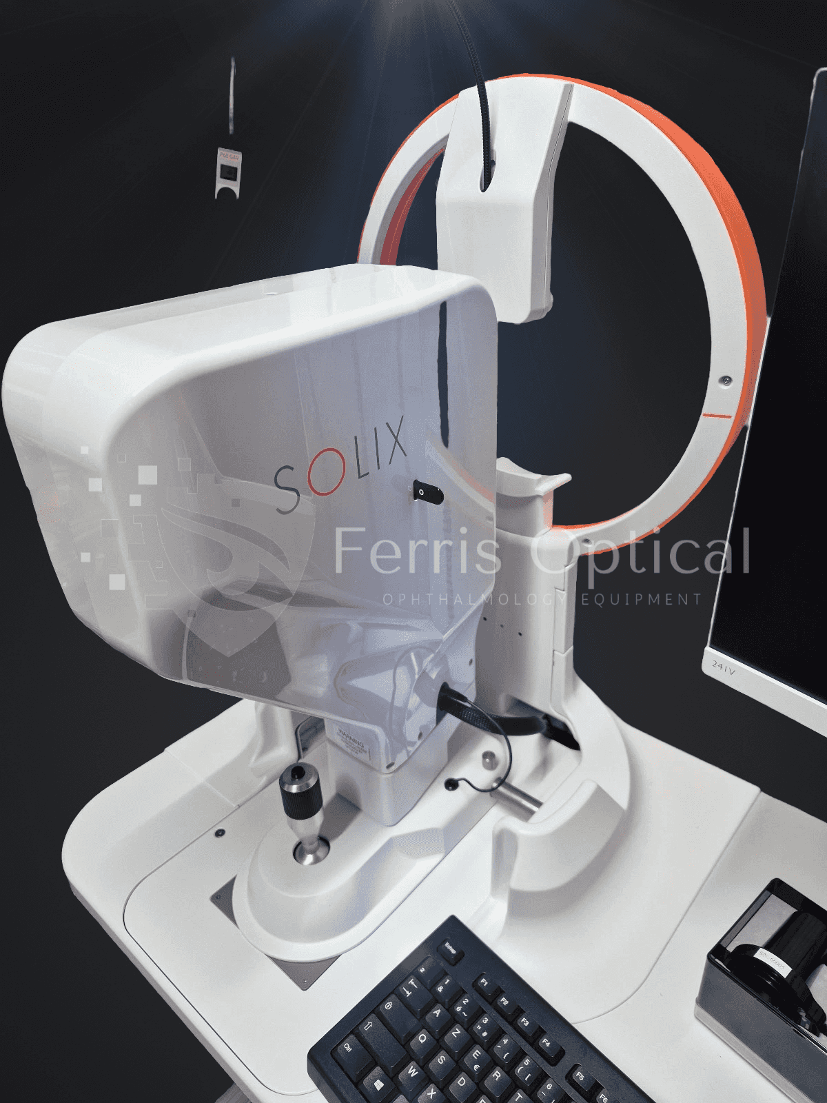
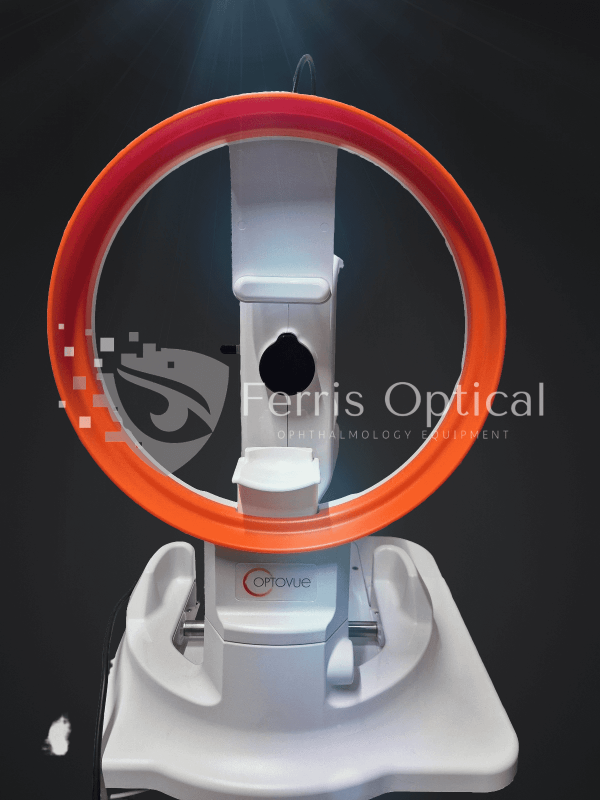
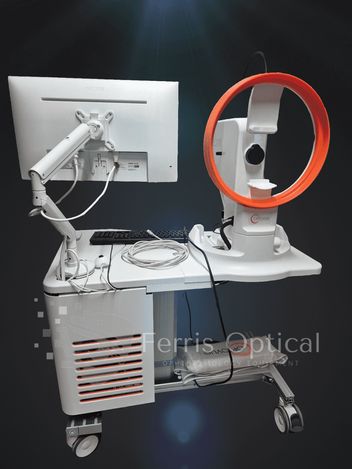
SOLIX es la tecnología innovadora que los optometristas necesitan para mejorar la atención al paciente y mantenerse a la vanguardia de la competencia. Construida sobre OCT de Dominio Espectral de alta velocidad, la plataforma FullRange® equipa a su clínica para identificar y tratar un amplio espectro de afecciones oculares, desde la córnea hasta la retina. Con SOLIX, proporcionará diagnósticos más rápidos y precisos, y ofrecerá una experiencia superior al paciente, diferenciando su clínica.
El Optovue Solix permite a los optometristas identificar anomalías retinianas detectadas en la fotografía del fondo de ojo, incluidas imágenes de campo amplio, y ver instantáneamente secciones transversales detalladas con OCT. Con escaneos ampliados de 6 mm por 6 mm del nervio óptico, su clínica obtiene mayor densidad de imágenes y datos de diagnóstico más nítidos y confiables.
El módulo de topografía avanzada ofrece las mediciones corneales precisas que los optometristas necesitan para un ajuste preciso de lentes de contacto y la planificación de cirugías refractivas. Detecta el queratocono de forma temprana con confianza, gracias a un análisis epitelial detallado. Realiza evaluaciones corneales anteriores y posteriores completas, distinguiendo las células epiteliales y el estroma, y monitoriza la curación y los resultados refractivos, todo a partir de un solo escaneo.
Informes personalizables con hasta 6 mapas simultáneos le brindan la información necesaria para tomar decisiones clínicas más inteligentes e impresionar a sus pacientes con tecnología que ofrece resultados.
Maximizar la utilidad clínica
Optovue Solix es la solución de próxima generación para clínicas de optometría listas para liderar en el cuidado oftalmológico. Con FullRange® OCT y OCT-Angiografía, una potente cámara de fondo integrada y una imagen IR externa, Solix ofrece la versatilidad y precisión de imagen que su clínica necesita. Su velocísima velocidad de escaneo de 120 kHz, patrones de escaneo densos y funciones de informes avanzados le permiten diagnosticar con mayor confianza, tratar de forma más proactiva y brindar el más alto nivel de atención a sus pacientes.
Detección de glaucoma
El Optovue Solix lleva la detección del glaucoma al siguiente nivel con sus avanzadas capacidades de imagen y análisis. Al incorporar herramientas sofisticadas como Dual Track, SSADA, MCT y segmentación de IA, el dispositivo proporciona un enfoque integral para la evaluación del glaucoma.
Estas tecnologías permiten a Solix ofrecer una reproducibilidad y repetibilidad excepcionales, con un rendimiento el doble de eficaz que los modelos anteriores. Esto garantiza un seguimiento fiable de la progresión de la enfermedad y la respuesta al tratamiento. Las funciones de imagen integradas permiten una visualización detallada de los cambios estructurales y vasculares asociados al glaucoma, proporcionando una visión holística de la afección.
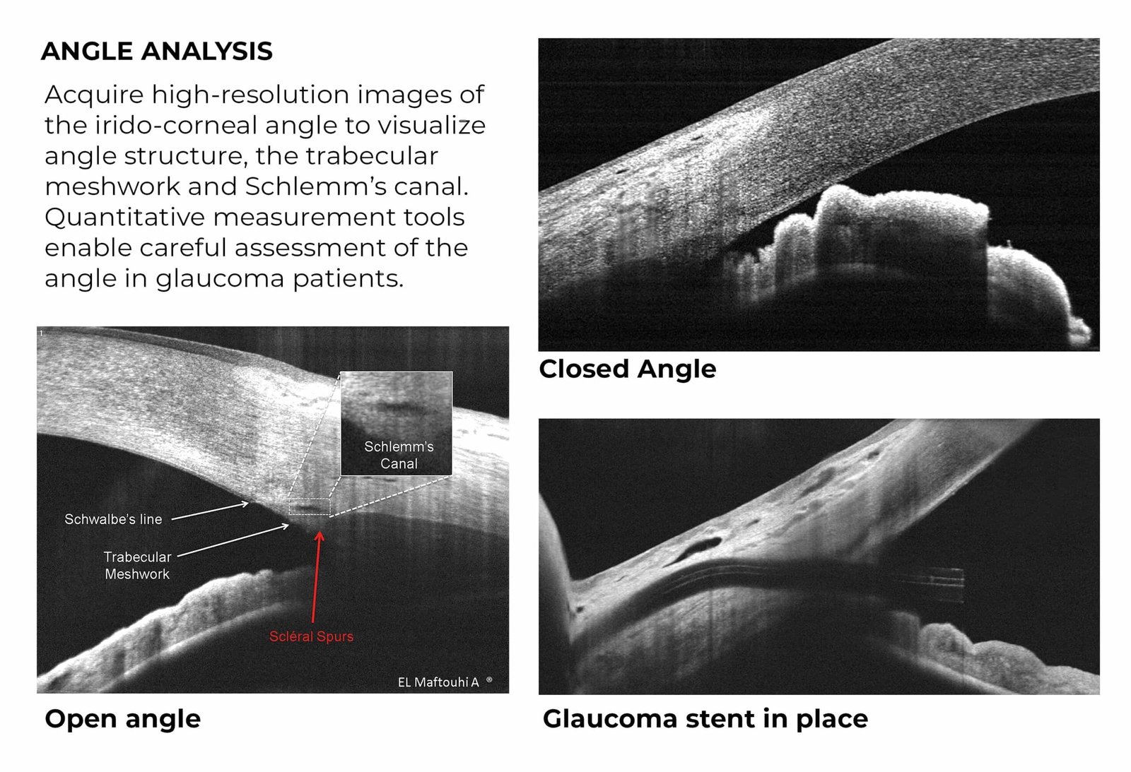
Las herramientas de medición cuantitativa permiten la evaluación precisa del ángulo iridocorneal, la malla trabecular y el canal de Schlemm, facilitando la detección temprana y el manejo continuo del glaucoma. Esta capacidad diagnóstica avanzada permite a los médicos tomar decisiones bien informadas.
FullRange® Retina
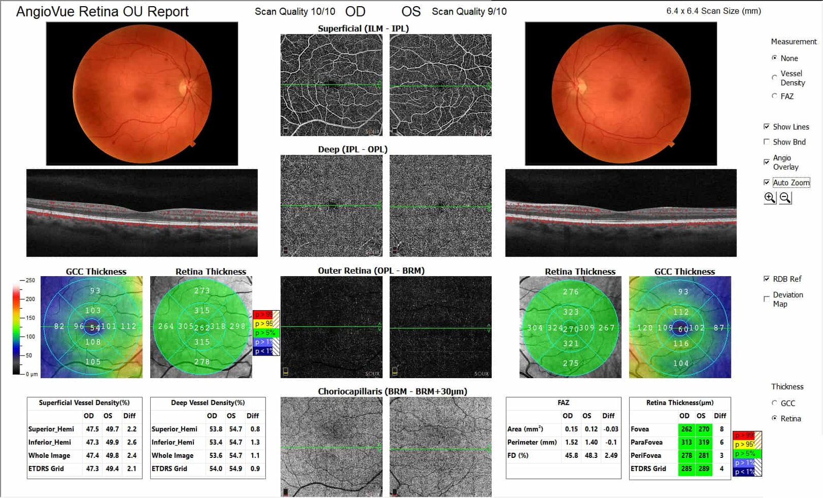
La función FullRange® Retina del Optovue Solix proporciona capacidades de imagen y análisis incomparables, incluso para pacientes con alta miopía. El dispositivo genera todas las imágenes y datos necesarios para una evaluación retiniana completa, asegurando que ningún detalle pase desapercibido.
La tecnología AngioVue OCTA ofrece una visualización y cuantificación precisas de la vasculatura retiniana, lo cual es crucial para diagnosticar y monitorizar una variedad de afecciones retinianas. La función Quad-Montage combina múltiples escaneos para obtener una visualización de campo amplio de la retina periférica, permitiendo un examen detallado de las patologías retinianas.
Adicionalmente, la renderización 3D OCTA permite la visualización de la conectividad vascular, proporcionando información sobre redes vasculares complejas. Estas capacidades de imagen integrales mejoran la capacidad para diagnosticar, monitorear y tratar enfermedades retinianas.
Bienestar y AngioBienestar
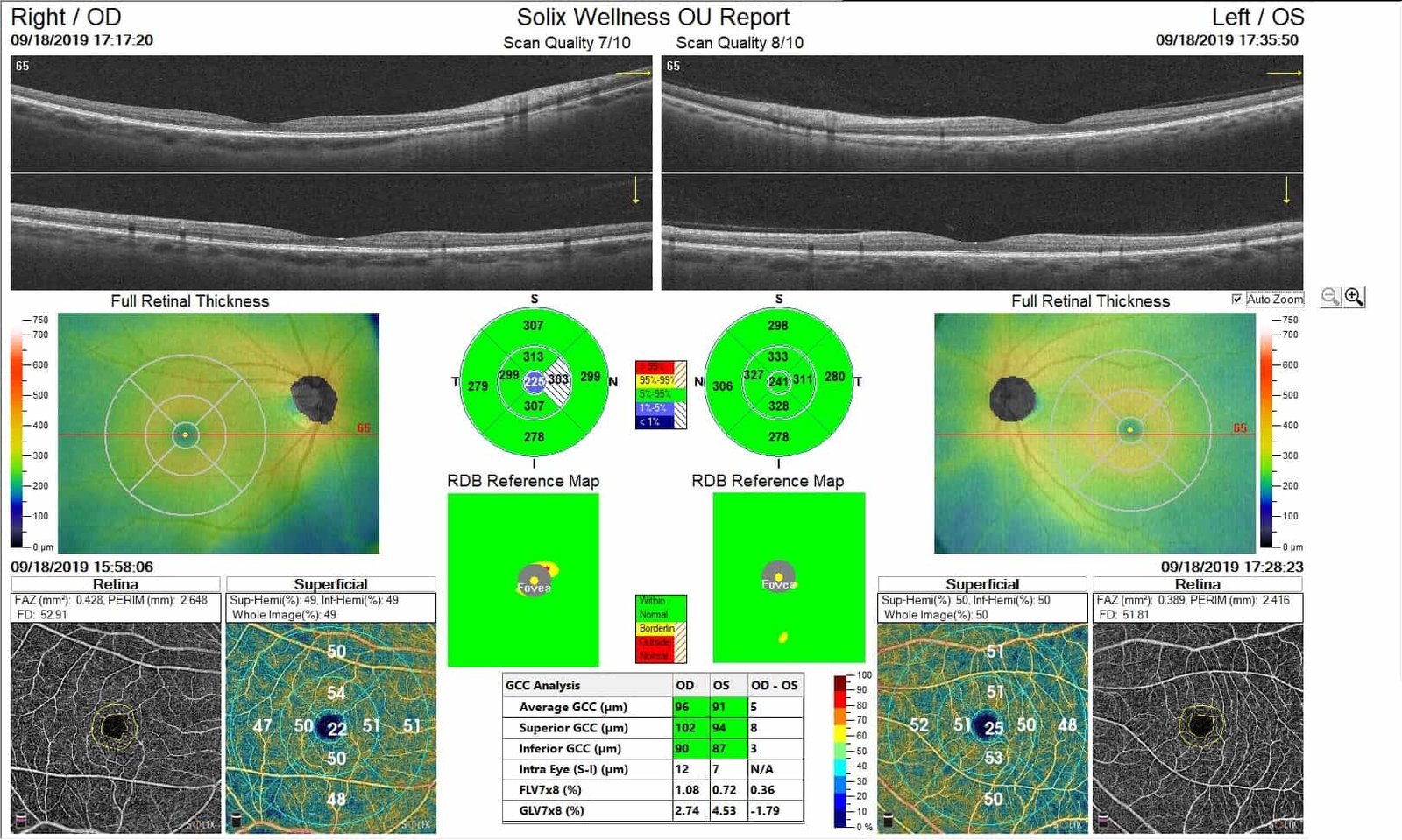
Las funciones Wellness y AngioWellness del Optovue Solix representan un nuevo estándar en la atención al paciente, particularmente para aquellos con sospecha de patologías retinianas y glaucoma. Estas capacidades agilizan el proceso de examen al integrar evaluaciones estructurales y vasculares en un único informe integral.
El escaneo AngioWellness proporciona una evaluación exhaustiva de pacientes diabéticos y sospechosos de glaucoma al combinar métricas sobre el grosor de la retina y de las células ganglionares con evaluaciones objetivas de la vasculatura retiniana. Este enfoque holístico descubre indicadores tempranos de cambios diabéticos y ayuda en la detección y manejo tempranos del glaucoma. Al ofrecer una visión detallada de la salud ocular del paciente, estos protocolos de bienestar mejoran la participación, retención y diferenciación de la práctica del paciente.
Referencia
 English
 Spanish
 German
 French