
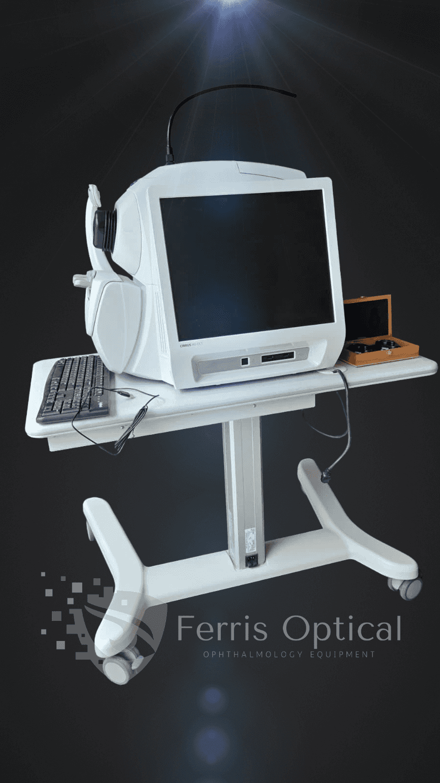
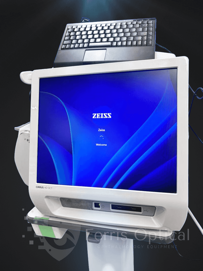
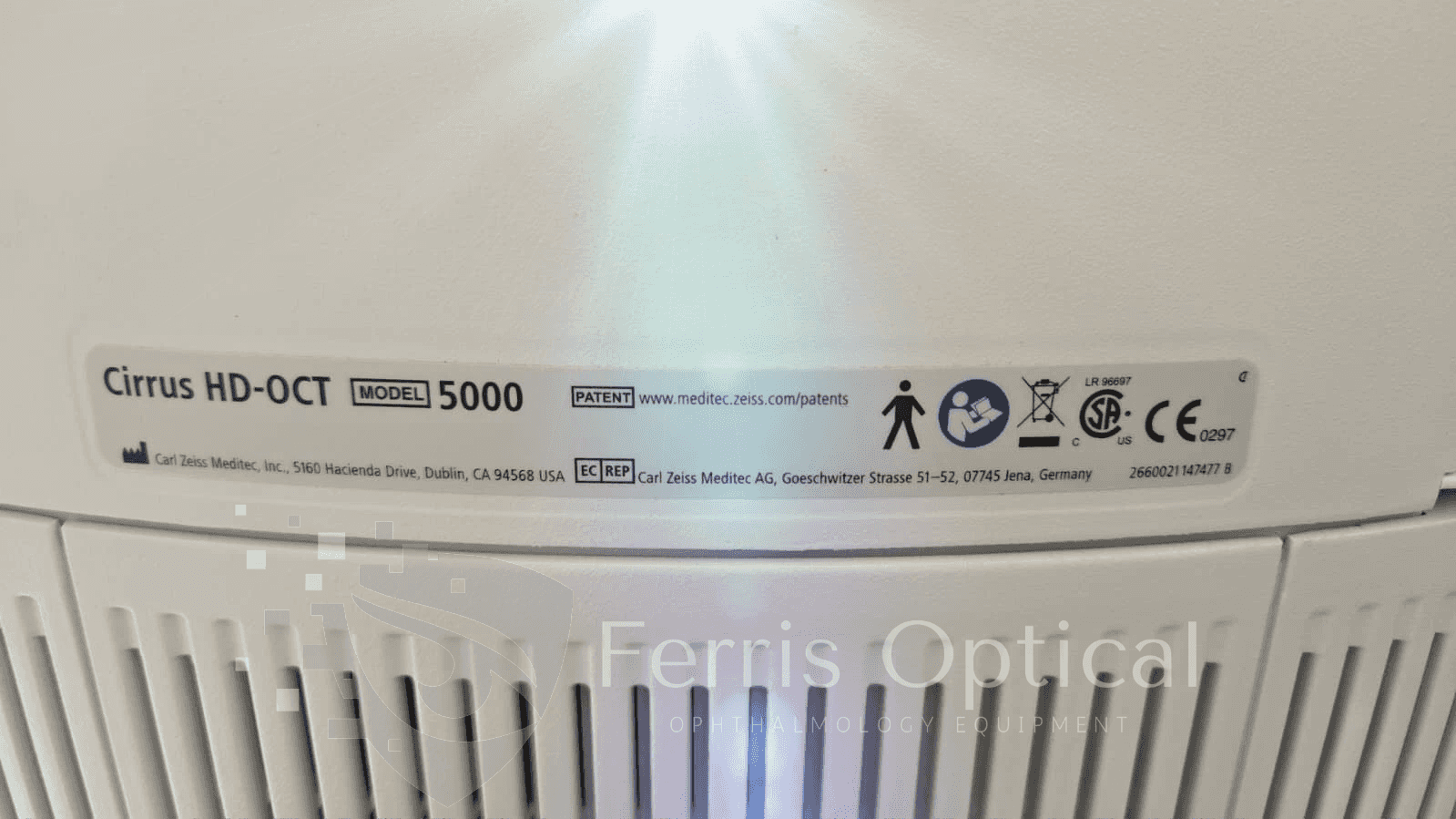
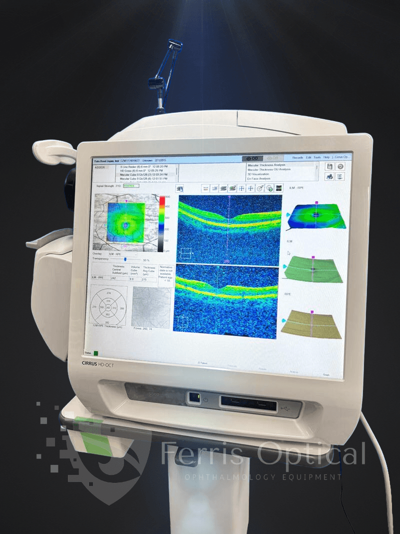
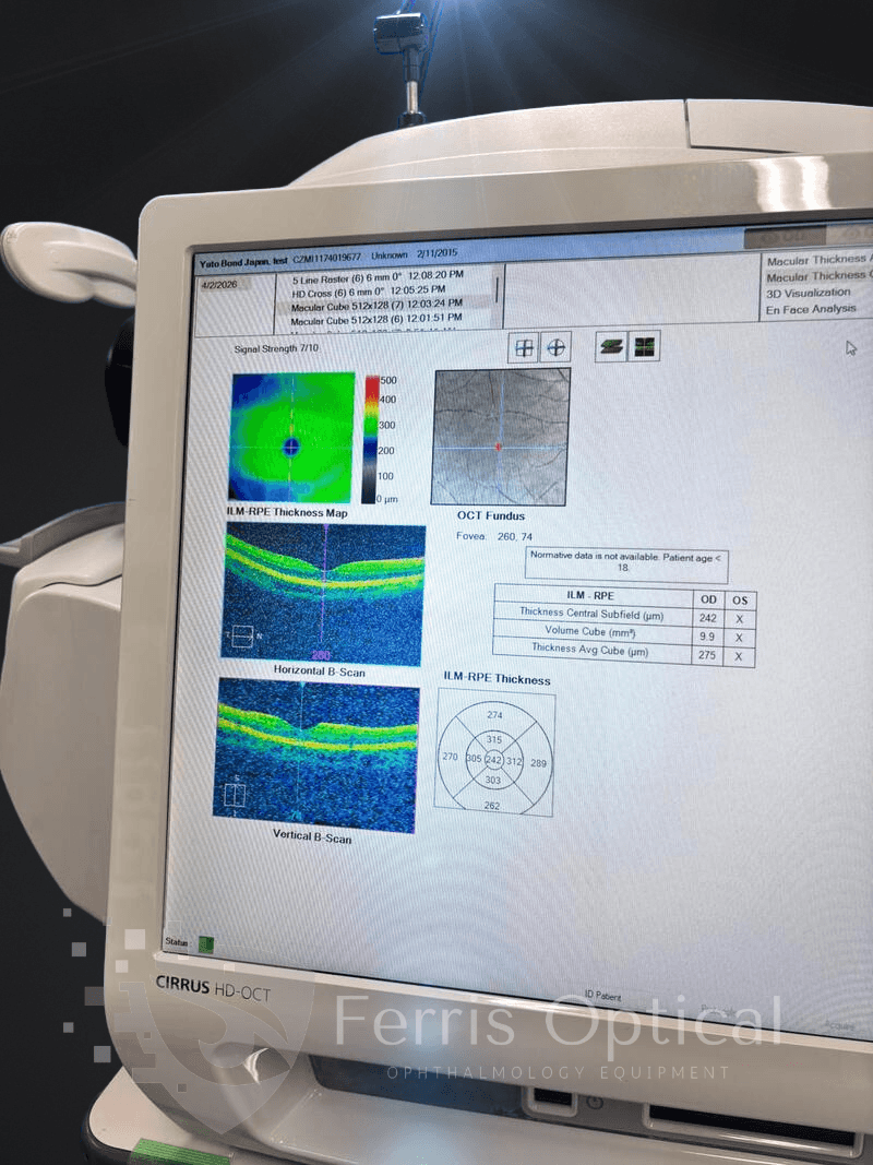
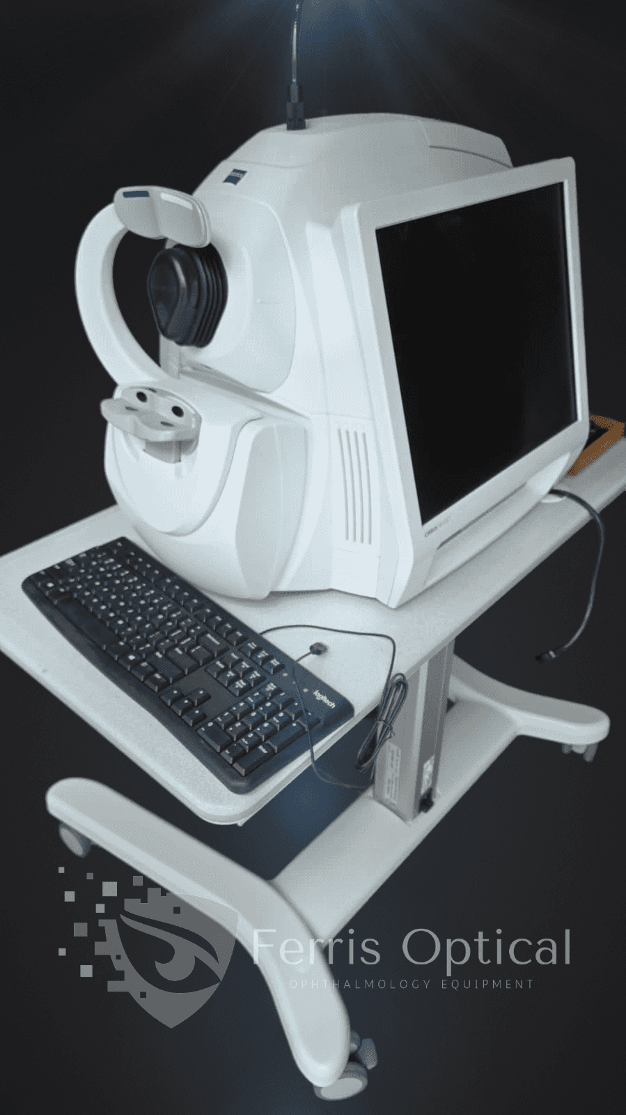
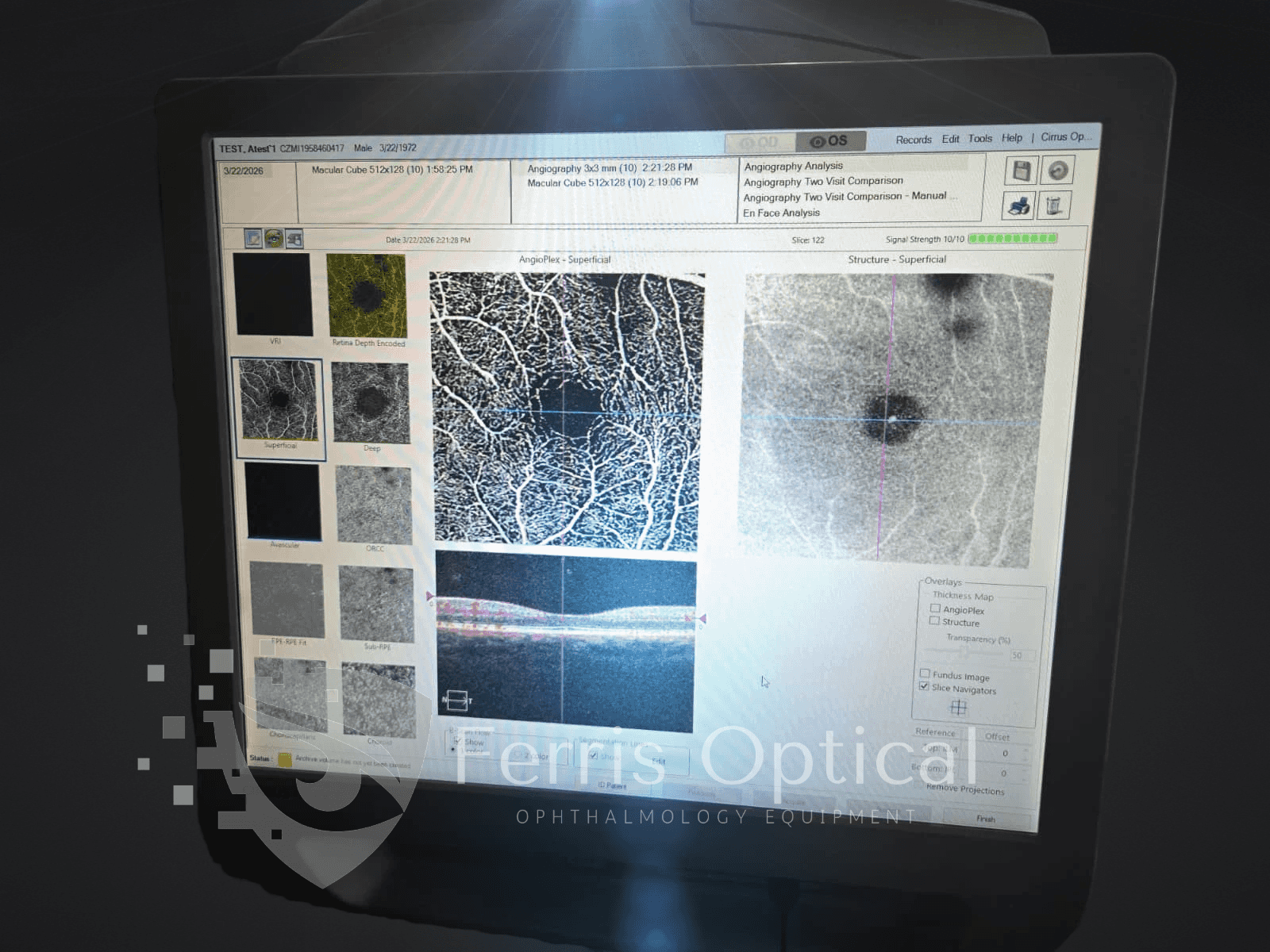
The ZEISS CIRRUS 5000 with AngioPlex is the perfect solution for optometry clinics seeking advanced, non-invasive retinal diagnostics. This system combines high-resolution OCT imaging with OCT Angiography (OCT-A), providing optometrists with detailed structural and microvascular analysis on a single, powerful platform—helping you detect, monitor, and manage retinal disease with confidence.
With the ZEISS CIRRUS 5000, optometrists get the highest resolution visualization and advanced clinical tools. Track retinal pigment epithelium integrity with confidence, assess subtle glaucomatous changes in the macula using Ganglion Cell Analysis, and reduce eye motion artifacts thanks to FastTrac™ retinal tracking. This proprietary scan strategy ensures you capture consistent, high-resolution images from visit to visit—critical for accurate disease progression monitoring and confident patient management.
Características clave
- * AngioPlex → non-invasive vascular imaging
- * FastTrac™ Eye Tracking → improved scan consistency
- * Advanced RPE Analysis → enhanced retinal assessment
- * Automatic follow-up comparison between visits
Zeiss Cirrus 500 OCT — Refurbished Spectral-Domain OCT for Optometrists: GPA and Ganglion Cell Analysis
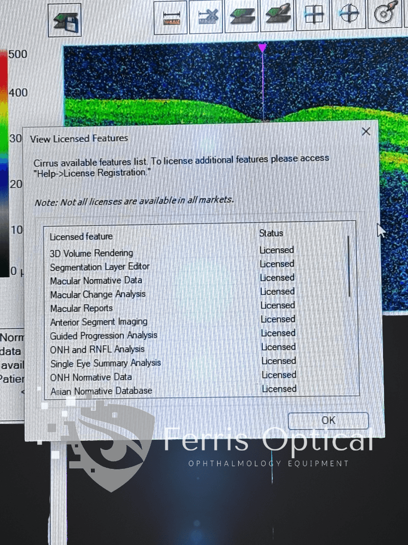
The Zeiss Cirrus 500 OCT is an ideal choice for optometrists seeking a reliable, clinically proven SD-OCT at a practical price. With Guided Progression Analysis (GPA), Ganglion Cell Analysis (GCA), and the full suite of Cirrus macular and optic disc protocols, optometry clinics can confidently track disease progression and compare results against robust normative data.
This platform is trusted by eye care professionals worldwide and supports all posterior segment imaging needs—from RNFL mapping and optic disc analysis to macular thickness and high-definition raster scans.
Peer-reviewed research validates that guided progression analysis on the Cirrus 500 OCT enables statistically accurate detection of glaucoma progression at every stage—empowering optometrists to deliver evidence-based care and meet co-management reporting standards.
Why Optometry Clinics Choose the Zeiss Cirrus 500 OCT
- * Guided Progression Analysis (GPA)—automatically detects RNFL thinning between visits and generates easy-to-interpret summary reports, making it the gold standard for glaucoma progression documentation in optometry clinics.
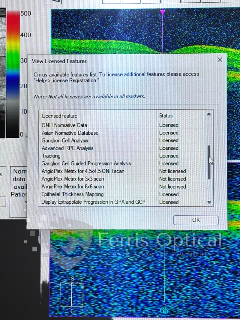
- * Ganglion Cell Analysis (GCA)—maps macular ganglion cells and inner plexiform layer, allowing optometrists to detect early glaucoma changes even before RNFL loss is visible.
- * 5-micron axial resolution at 840 nm—delivers crisp, detailed images for precise RNFL thickness measurement, macular mapping, and structural AMD monitoring in the optometry setting.
- * Full Cirrus protocol suite—comprehensive macular, optic disc, and anterior segment protocols are built in, supporting all standard clinical workflows for optometry clinics.
- * Cirrus normative database—age-stratified RNFL and GCC reference values provide optometrists with confidence in every diagnostic comparison.
- * Anterior segment capable—add the optional lens to image the cornea, angle, and anterior chamber in your optometry clinic without extra equipment.
- * Familiar Cirrus interface—enjoy a consistent user experience and minimal learning curve for optometrists already familiar with Cirrus systems.
Aplicaciones clínicas para optometristas
- * Glaucoma progression monitoring — GPA identifies statistically significant RNFL changes between visits; supported by the Cirrus normative database validated across hundreds of peer-reviewed glaucoma studies
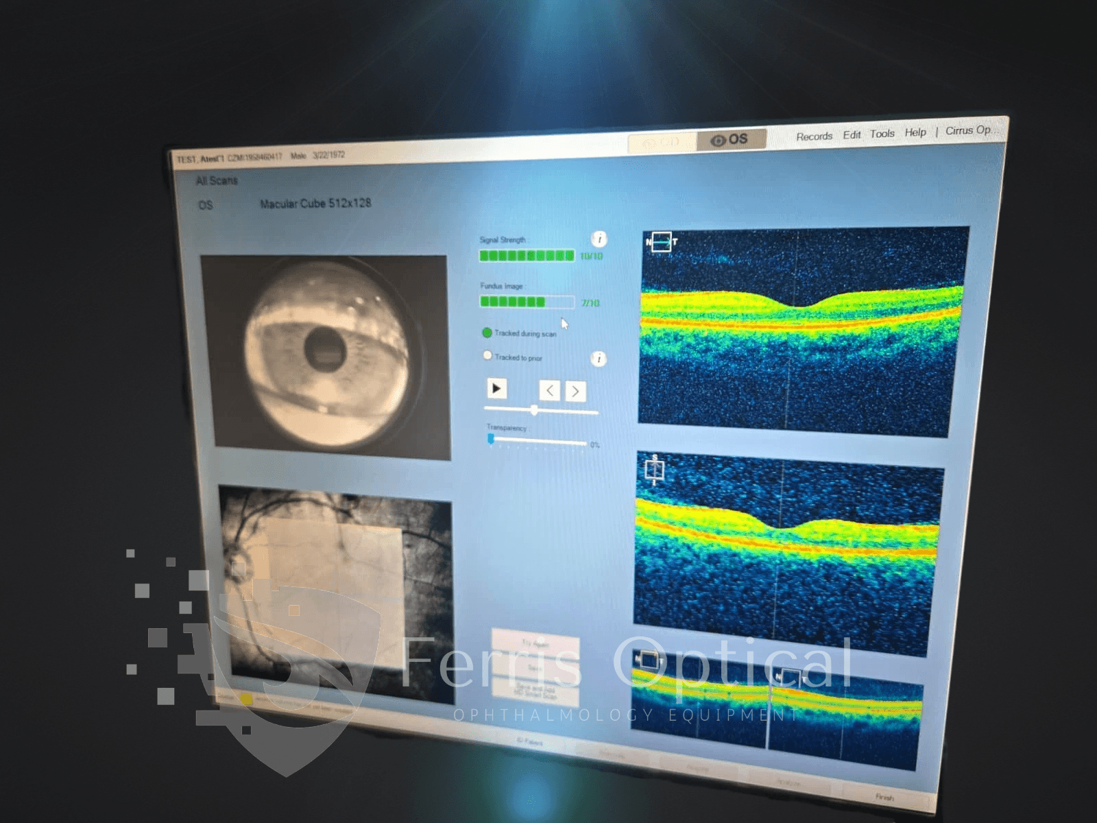
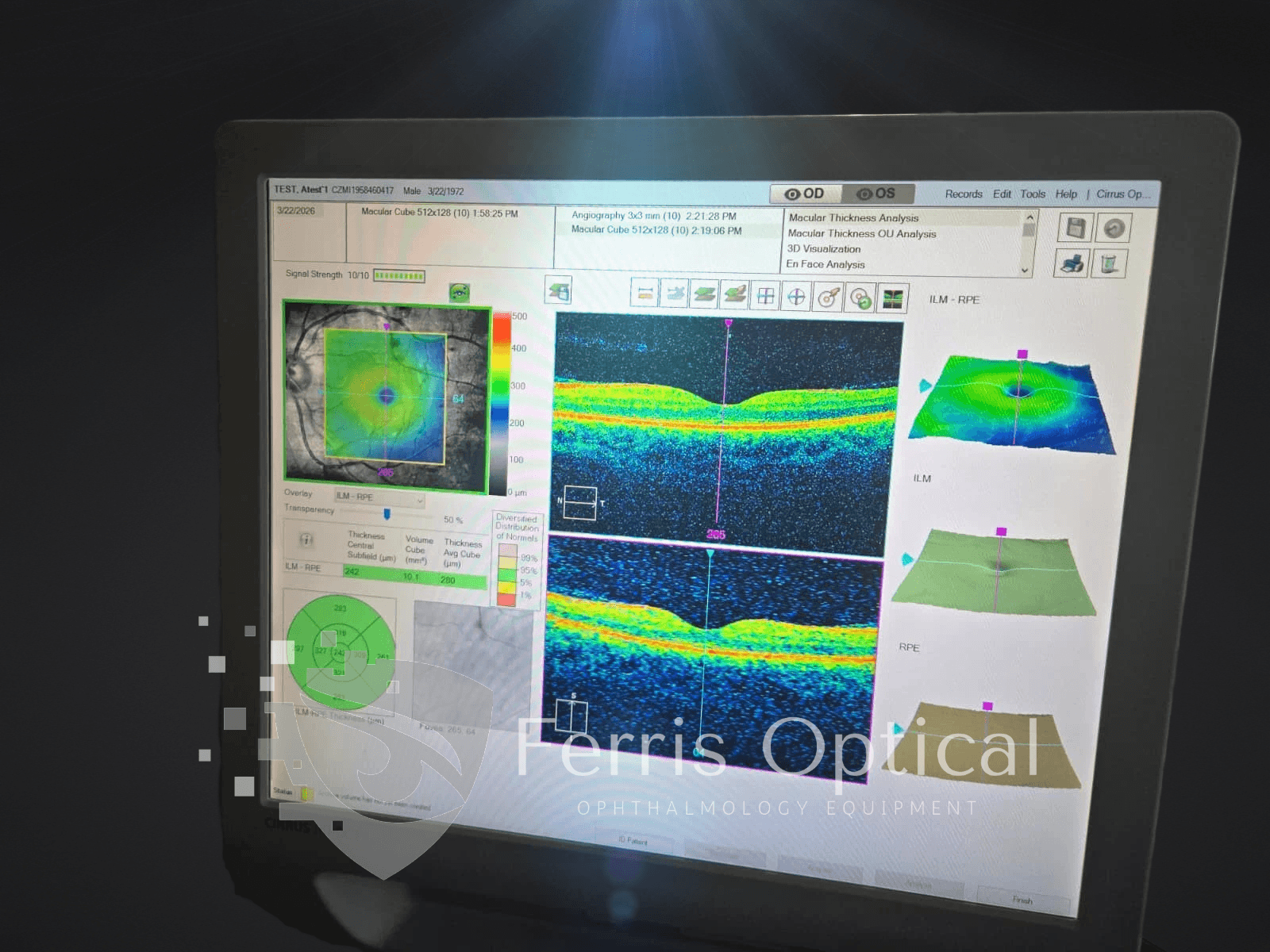
- * Macular disease surveillance — AMD, DME, epiretinal membrane, and vascular occlusion imaging with macular cube acquisitions and thickness maps for longitudinal follow-up
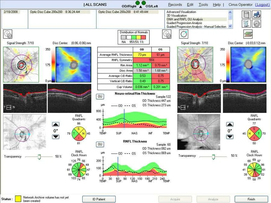
- * Optic disc documentation — Optic Disc Cube 200×200 captures the full disc and peripapillary RNFL in a single acquisition; disc parameters for baseline and follow-up reporting
- * Pre- and post-operative evaluation — macular thickness maps and RNFL status for cataract, vitreoretinal, and glaucoma surgical planning and outcomes documentation
- * Anterior segment imaging — corneal and anterior chamber assessment using the optional anterior segment lens
¿Quién se beneficia en optometría?
- * Glaucoma specialists — GPA and GCA provide the statistical rigor and documentation required for progression monitoring and managed care reporting
- * Comprehensive ophthalmology and optometry practices — single instrument covers RNFL analysis, macular screening, and optic disc documentation for the full patient panel
- * Practices upgrading from older OCT platforms — the Cirrus normative database and GPA toolset represent a meaningful clinical step up from first-generation time-domain instruments
- * Optometrists entering glaucoma co-management — the Cirrus platform is accepted by the major co-management reporting frameworks; GPA provides the change documentation required
- * Multi-location groups standardizing on Cirrus — consistent platform, protocols, and normative values across all sites with the Cirrus 500
** WHY OPTOMETRISTS CHOOSE CIRRUS 5000 WITH ANGIOPLEX
- * Structural + vascular imaging in one system
- * Detection of microvascular changes
- * Reliable RNFL & Ganglion Cell Analysis (GCA)
- * Consistent follow-up and progression tracking—ensure accurate, repeatable scans for long-term patient management and co-management referrals.
**CONFIGURATION
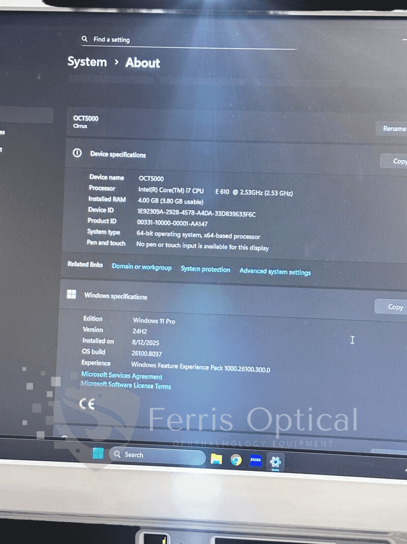
- Windows 10 system
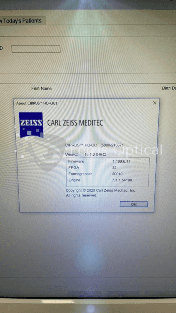
- Software Version: 11.5.3
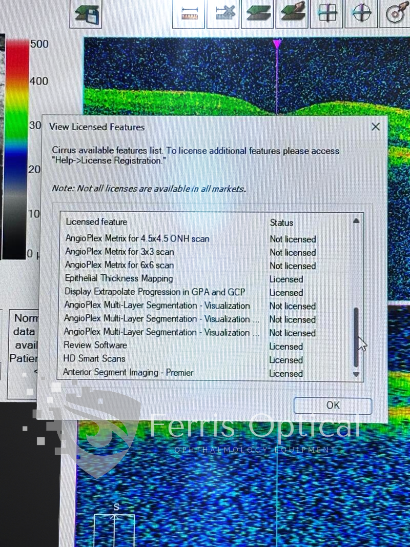
- Licenses: All activated
- AngioPlex OCT Angiography (OCT-A) enabled
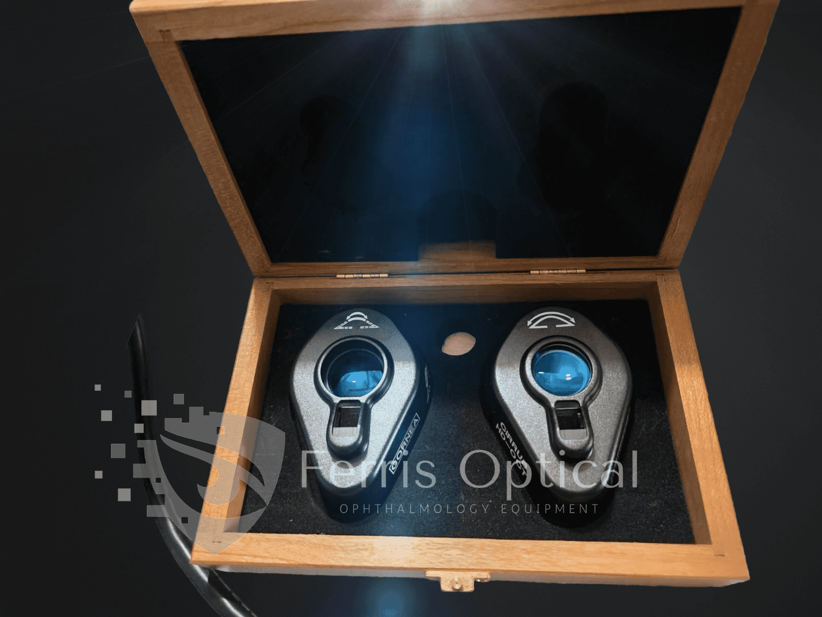
- Anterior Segment Premier Lens
** CONDITION
- • Fully functional — tested and verified
- • Professionally refurbished
- • Excellent cosmetic condition
- • Ready for immediate clinical use
** WHAT’S INCLUDED
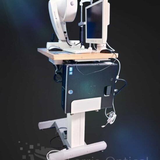
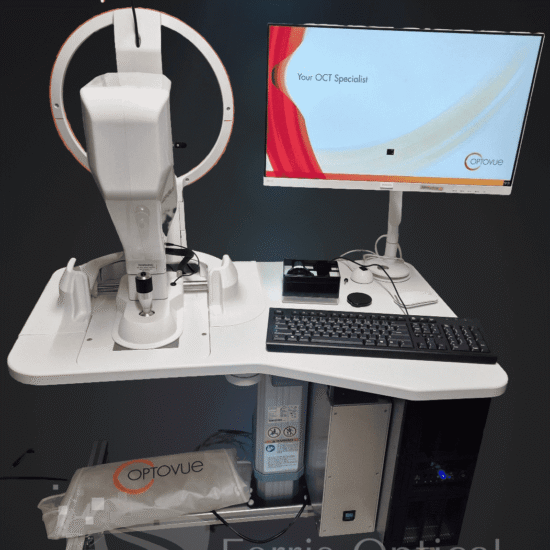
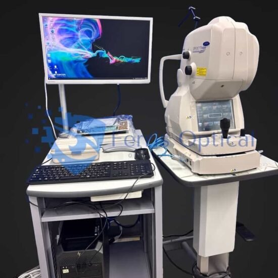
- * Zeiss Cirrus HD-OCT 500 main unit
- * Integrated workstation with Cirrus software
- * Patient chin and forehead rest assembly
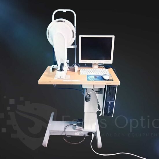
- * Monitor, keyboard, and mouse
- * Power and data cables
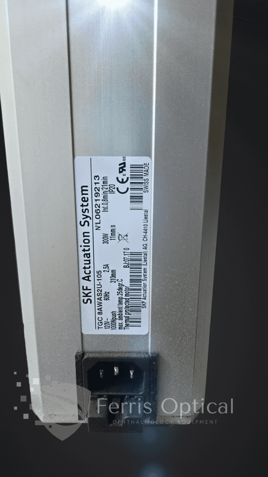
- * Electric table
- * 6-month warranty
- * Windows 10 System (integrated)
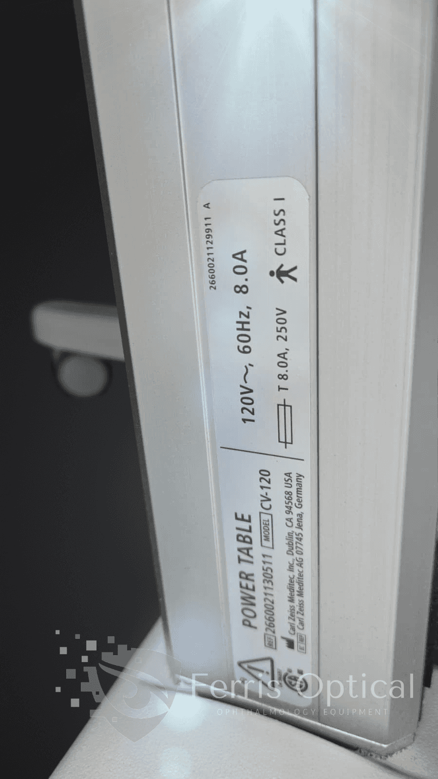
- * Power Table
- * User Manual
- * All cables and accessories
- * Anterior Premier lenses
- * Professional packaging
- * Remote Training
- * Remote Installation Support
- * Pre-purchase Consultation
- * Post-installation Guidance
Technical Specifications
| Spec | Detail |
| Manufacturer | Carl Zeiss Meditec |
| Model | Cirrus HD-OCT 500 |
| Technology | Spectral-domain OCT (SD-OCT) |
| Scan Speed | 27,000 A-scans/second (standard protocols) |
| Wavelength | 840 nm |
| Axial Resolution | 5 µm (tissue) |
| Scan Width | 6 mm × 6 mm |
| Imaging Depth | 2 mm |
| Protocols | Macular Cube 512×128, Optic Disc Cube 200×200, HD 5-Line Raster, anterior segment |
| Analysis Tools | GPA, GCA, RNFL thickness maps, optic disc parameters, macular thickness maps |
| OCTA | Not included (see Cirrus 5000 with AngioPlex) |
| Condition | Refurbished |
| Warranty | 6 months |
 English
 Spanish
 German
 French