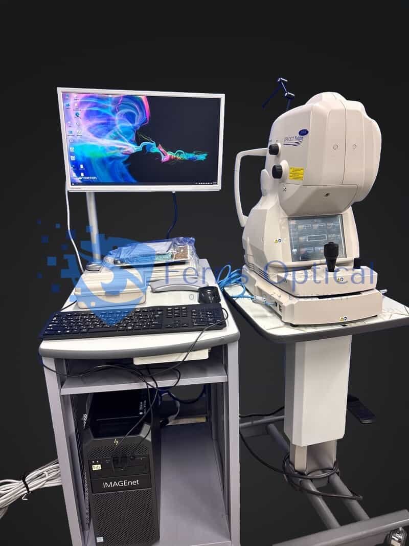
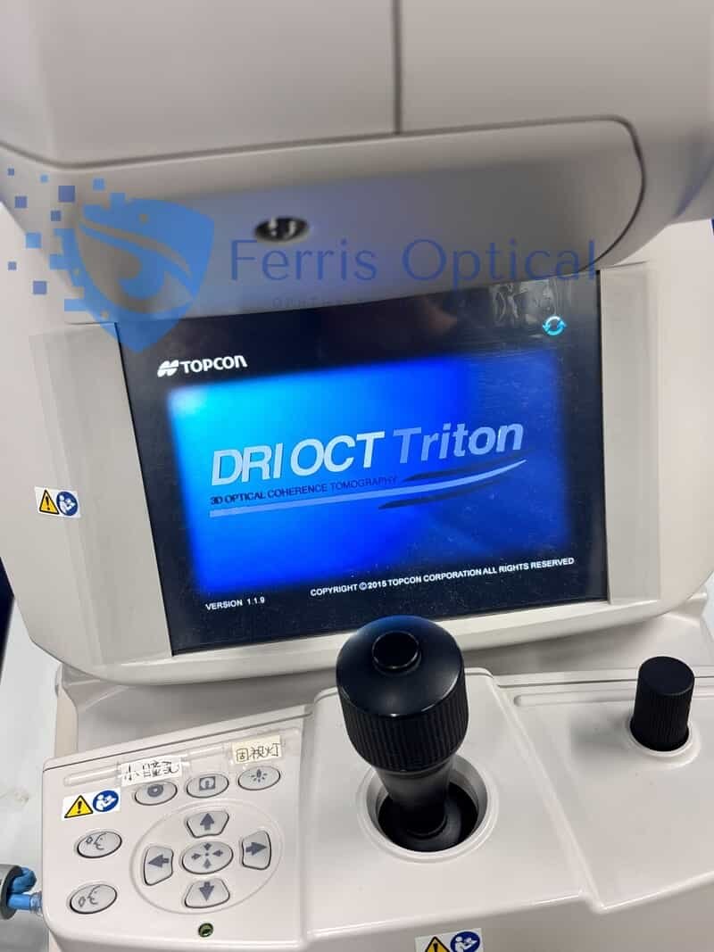
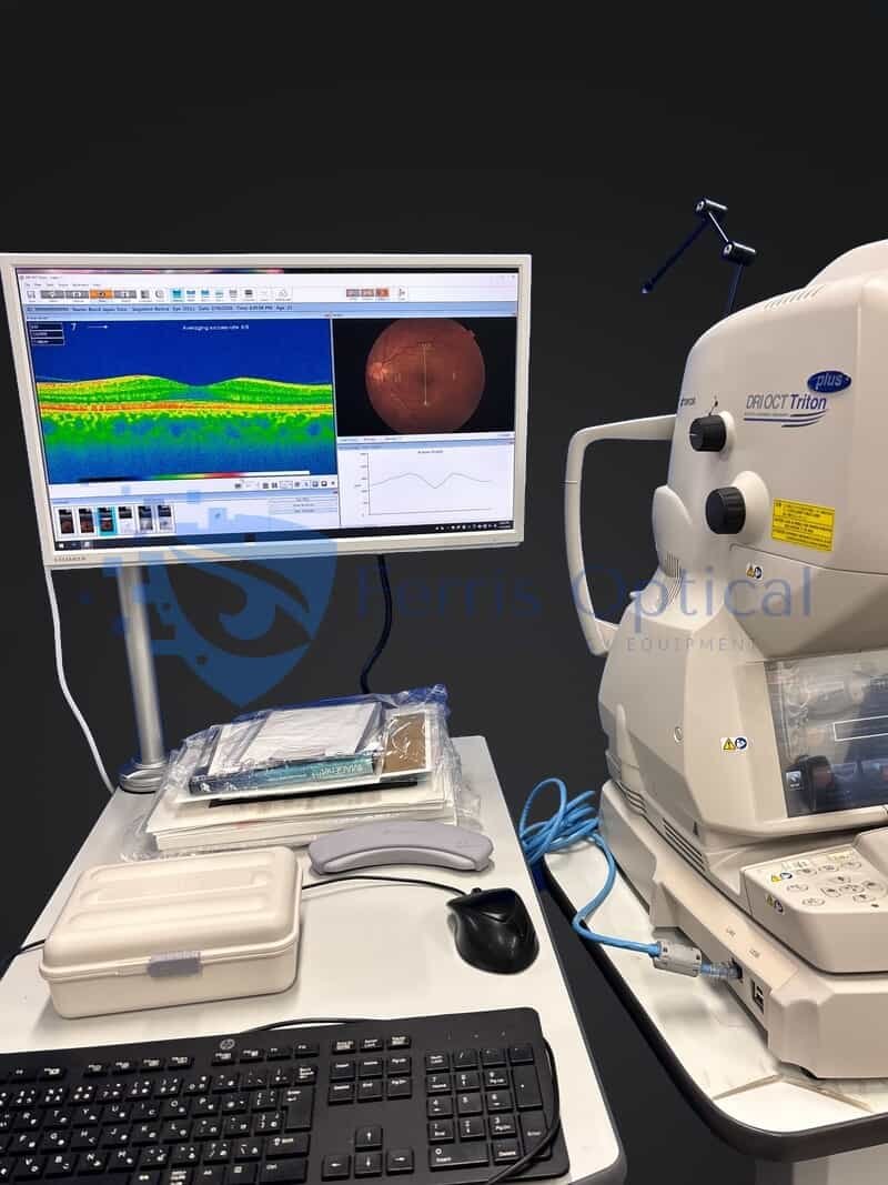
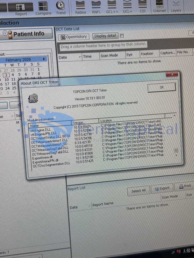
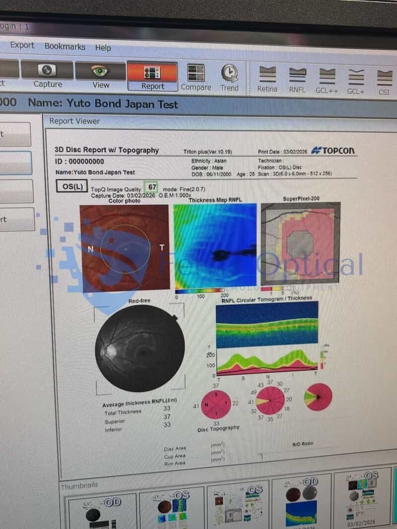
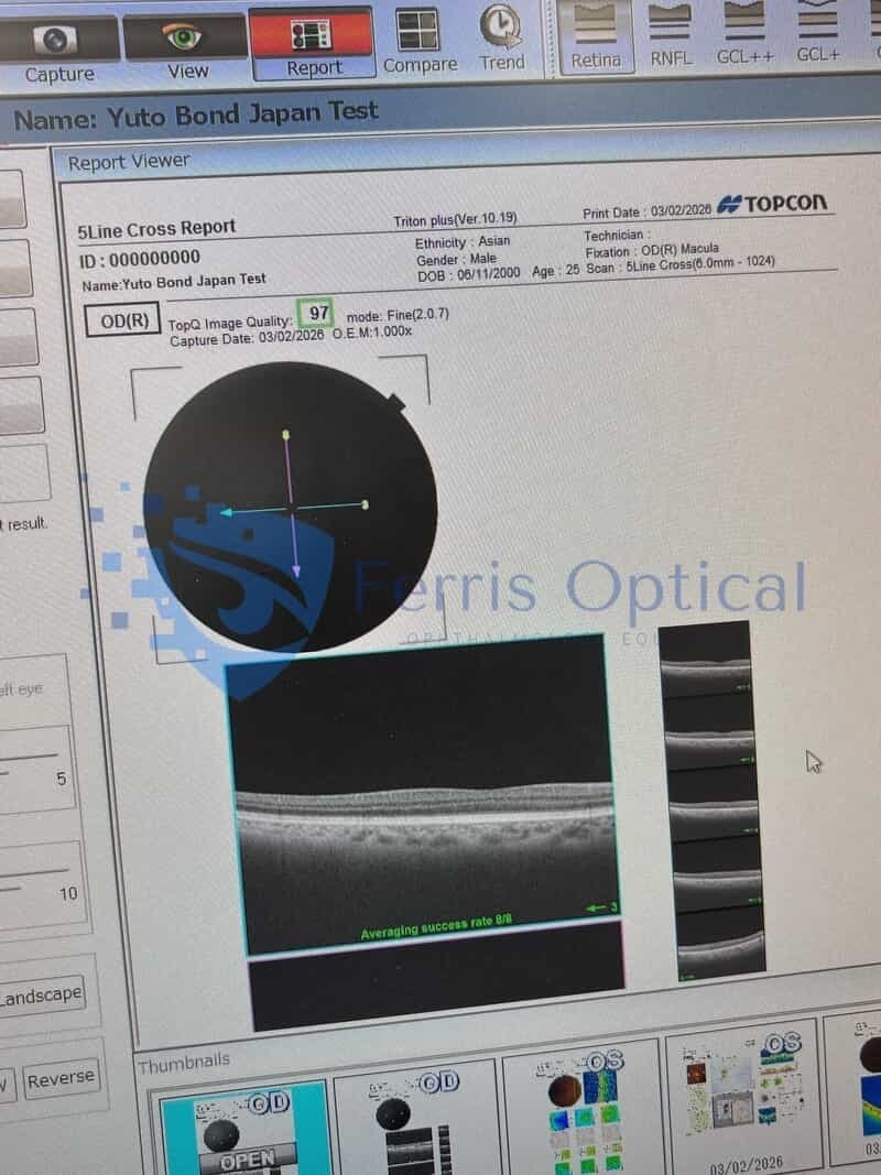
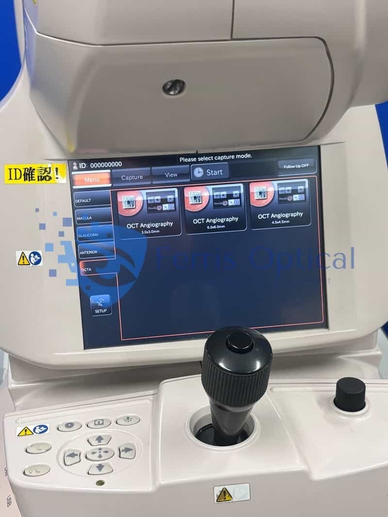
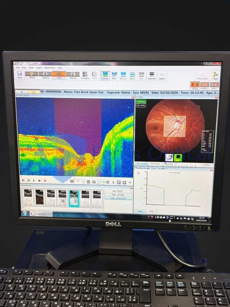
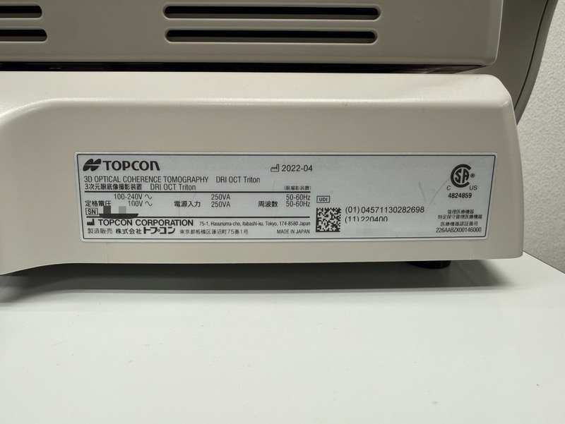
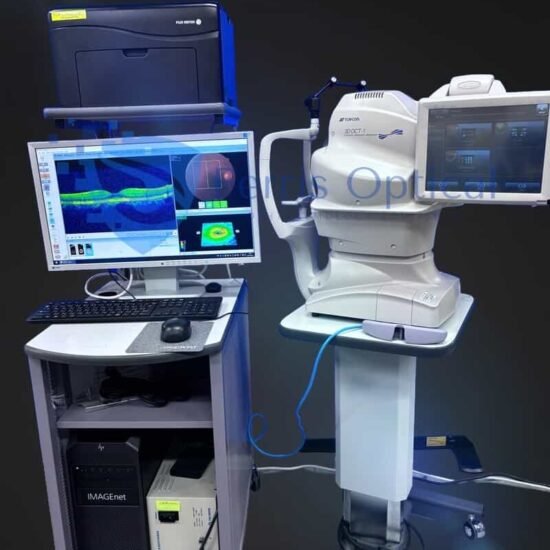
DRI OCT Triton Hauptmerkmale
- Swept-Source OCT mit einer Scan-Geschwindigkeit von 100.000 A-Scans/Sekunde
- 1050 nm Wellenlänge
- Multimodale Bildgebung: True-Color-Fundusfotografie, OCT-Angiographie(1), Weitfeld für OCT und OCT-A(1), Vorderabschnitt(1), FA*, FAF*
Bildqualität
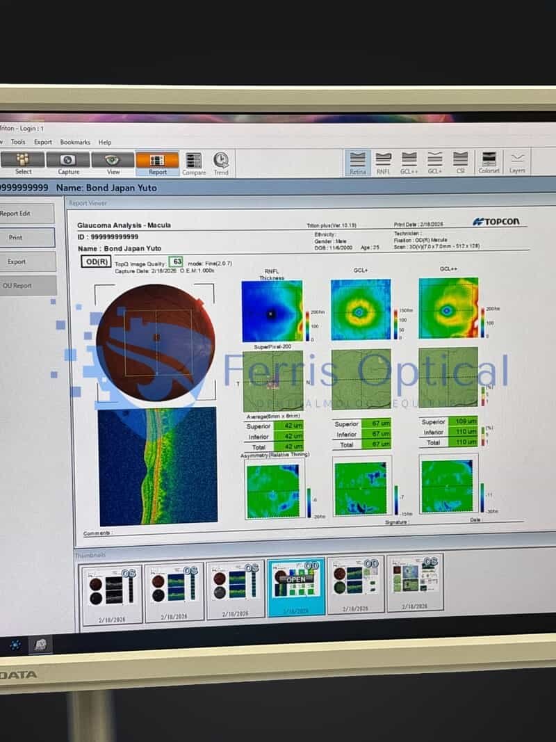
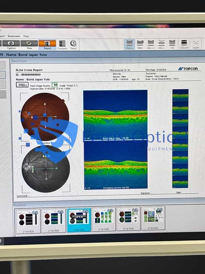
Das Swept-Source-OCT von Triton mit seiner Scangeschwindigkeit von 100 kHz und der Wellenlänge von 1.050 nm liefert selbst von den tiefsten Schichten des Auges klare und detaillierte Bilder mit kurzer Aufnahmezeit. Visualisieren Sie nicht nur Retina und Glaskörper, sondern auch Aderhaut und Sklera.
NEU: Smart Denoise entdecken1, Topcons einzigartiger KI-Algorithmus, der Artefakte reduziert und den Kontrast auf OCT und OCT-A erhöht1.
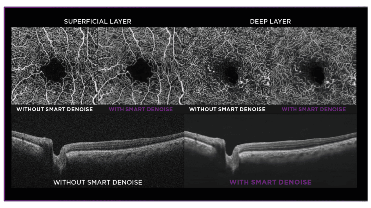
Diagnosefähigkeit
Tiefere Einblicke ermöglichen ein besseres Verständnis vieler Augenkrankheiten.2. Mit Funktionen wie OCT-Angiographie1, Fundusautofluoreszenz* und Vor OCT, Triton befähigt Kliniker mit multimodaler Bildgebungsfähigkeit zur Beurteilung und Erhaltung der Augengesundheit von Patienten.
Effizienz beim Üben
Die automatisierten Funktionen des Triton, einschließlich Einzelbildaufnahme und SMARTTrackTM Systeme, die entwickelt wurden, um Ihren.
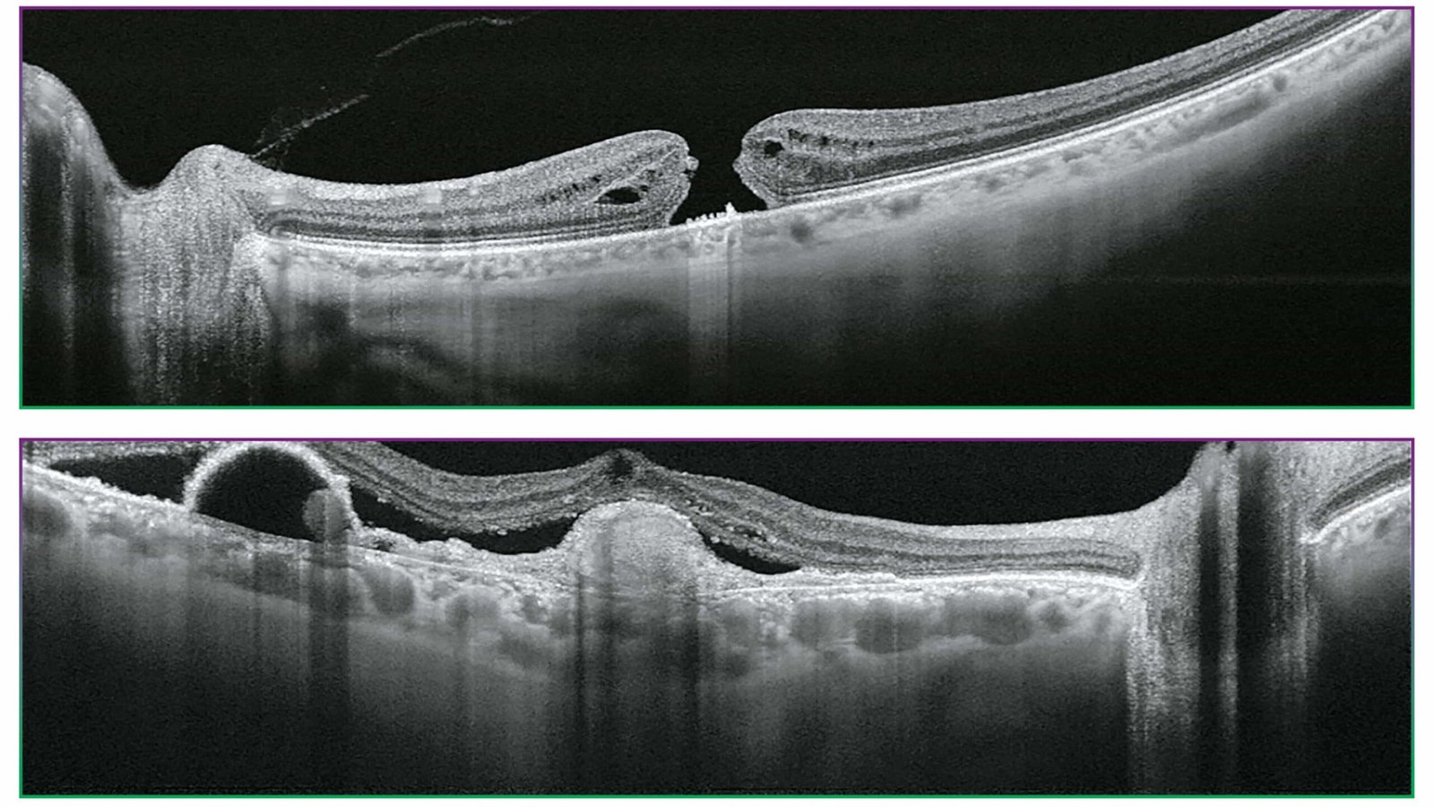
Swept-Source OCT Bildgebung
1.050nm Wellenlänge
Das längerwellige Licht bietet eine bessere Gewebepenetration und ermöglicht so die Visualisierung der tiefsten Schichten des Auges.2.
Swept-Source-OCT-Technologie und eine Scangeschwindigkeit von 100.000 A-Scans/Sek.
Die schnelle Scan-Geschwindigkeit von 100.000 A-Scans/Sekunde ermöglicht die Aufnahme klarer B-Scans3 durch den Erwerb von mehr A-Scans innerhalb einer gegebenen Bildaufnahmezeit. Dies hilft, Artefakte durch unfreiwillige Augenbewegungen wie Sakkaden und Blinzeln zu reduzieren.
Unsichtbare Scanlines
Das unsichtbare Licht mit einer Wellenlänge von 1.050 nm hilft Patienten, sich während des Scans auf das Fixationsziel zu konzentrieren, und reduziert so unfreiwillige Augenbewegungen. Es unterstützt einen effizienteren Arbeitsablauf in einer Praxis, da weniger erneute Scans erforderlich sind.
Swept-Source-OCT integriert multimodale Fundusbildgebung
Wahre Farbe6 Fundusbilder
Der DRI OCT Triton bietet ein echtes Farbbild des nicht mediatisierten Fundus. Fluoreszenzangiographie (FA)* und Fundus-Autofluoreszenz (FAF)* sind verfügbar, um die diagnostischen Fähigkeiten des Triton Plus zu erweitern.
Das All-in-One-Gerät unterstützt einen effizienten Arbeitsablauf in der Praxis. Swept Source OCT integriert multimodale Fundusbildgebung. DRI OCT Triton kann das OCT- und Fundusbild in einer einzigen Aufnahme erfassen.
PinPoint™ Registration identifiziert die Position des B-Scans auf dem Fundusbild. Der Vergleich zwischen B-Scan und Fundusbild kann die klinische Effizienz während der Diagnose unterstützen.
NEU: Weitfeld-OCT und OCT-A
Die optionale Weitwinkel-Vorsatzlinse ermöglicht die Erfassung von Scans mit einer Länge von bis zu 21 mm. Gewinnen Sie tiefere klinische Erkenntnisse mit OCT- und OCT-A-Visualisierung. Die Weitfeld-Bildgebung ist bei einer Vielzahl von Erkrankungen wertvoll.
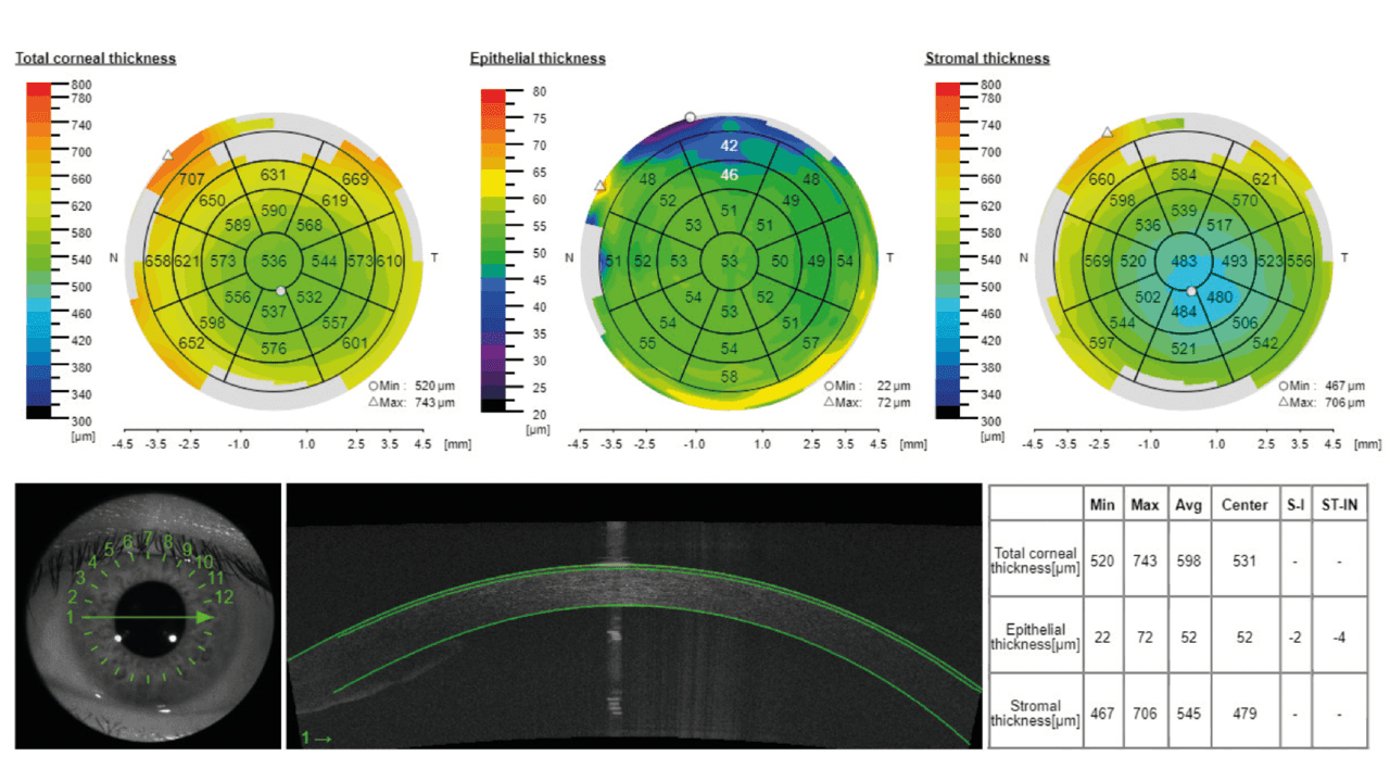
NEU: Vorderabschnittsmetriken
Die optionalen Vorderkammer-Bildgebungsfunktionen des Triton ermöglichen die Visualisierung von Hornhaut, Kammerwinkel, Iris und Vorder sclera und integrieren eine quantitative Analyse.
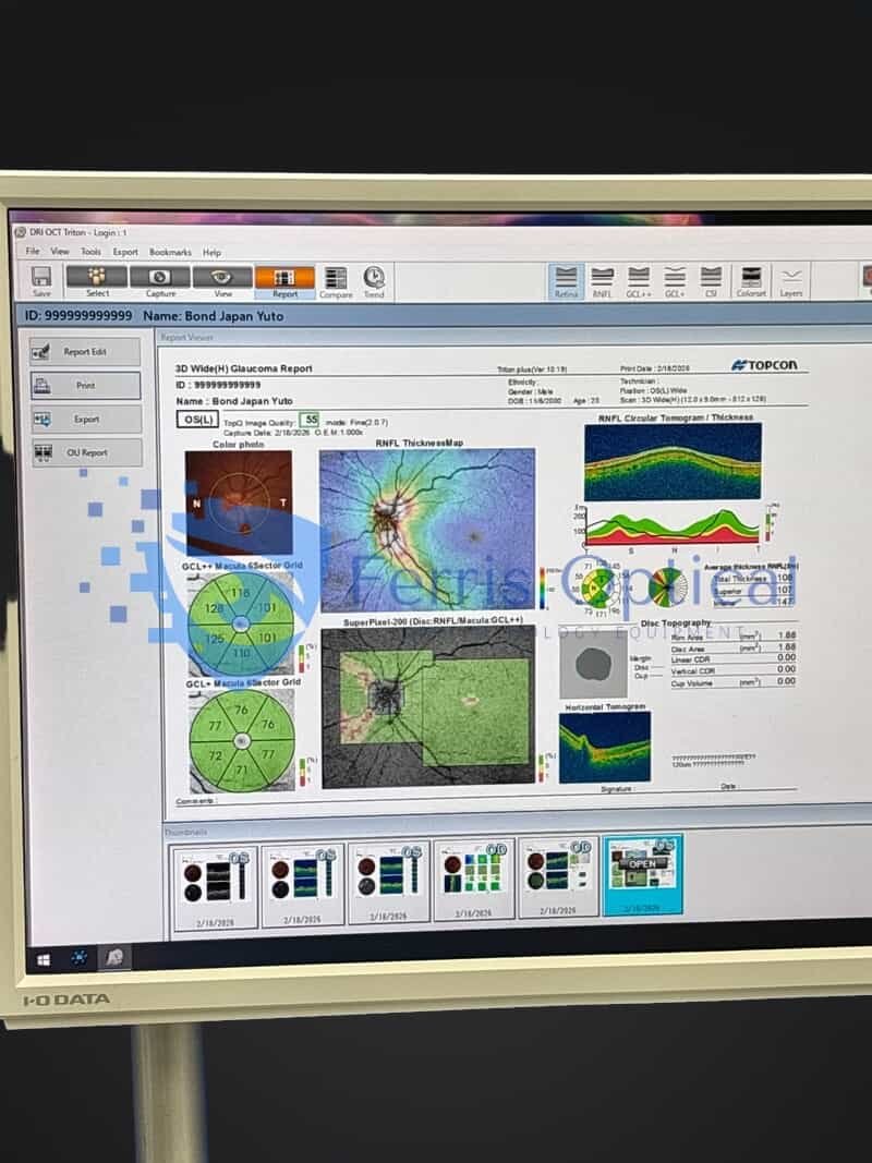
Die neue Vorderabschnittsfunktion bekräftigt den Wert von Triton in Augenheilanstalten mit detaillierten Berichten.
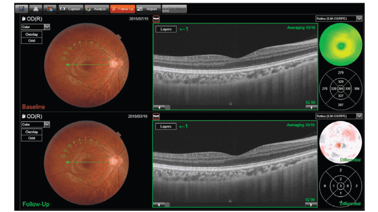
Follow-up-Funktion
Diese Funktion ermöglicht es, denselben Ort bei Nachuntersuchungen abzurufen und erneut zu analysieren, um vergangene und aktuelle Bilder zu vergleichen. Alles, was ein Bediener tun muss, ist einfach die vergangenen Daten auszuwählen, und Triton erfasst automatisch denselben Bereich.
Entdecken Sie weitere Möglichkeiten: Sehen Sie weiter und tiefer
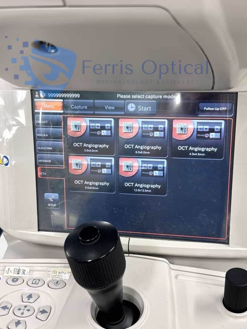
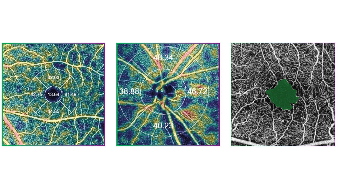
SS-OCT Angiographie
Topcons SS-OCT Angio™ kombiniert OCT-Angiographie mit Swept-Source-Technologie.
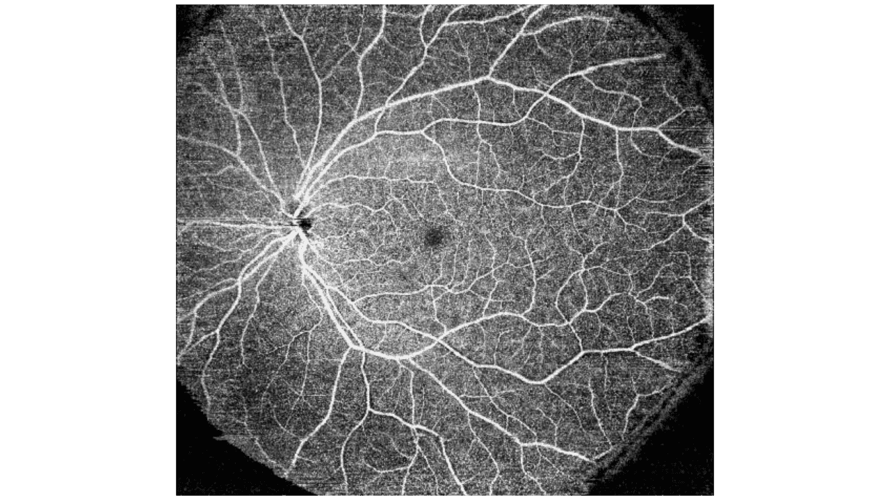
OCTARA™, ein proprietärer Bildverarbeitungsalgorithmus, bietet hochempfindliche angiografische Detektion4 welche die Darstellung von Gefäßstrukturen auch in der Aderhaut und den tieferen Netzhautschichten ermöglicht.
OktawaTM
OktawaTM ist die Bildverarbeitungstechnologie, die Signaländerungen extrahiert, die vom vaskulären Fluss herrühren, unter Verwendung mehrerer OCT-B-Scans, die an derselben Position aufgenommen wurden. Sie weist eine hohe Empfindlichkeit für die Erkennung auf
des geringen Blutflusses in der Mikrovaskulatur4.
Hochempfindliche Bildgebung und tiefere intravaskuläre Flussvisualisierung1
Dank der Swept-Source-Technologie und OCTARA™ lassen sich tiefere Strukturen mit einem geringeren, von der Tiefe abhängigen Signalabfall darstellen.3. Zusätzlich ermöglicht die Wellenlänge von 1 μm die OCT-Angiographie bei Patienten mit medialen Trübungen.
Schnelles Scannen, Echtzeit-Augenverfolgung
Bei 100.000 A-Scans pro Sekunde, gekoppelt mit unsichtbarer5 Mithilfe von Scanlinien und dem Eye-Tracking-System SMARTTrack™ erfasst Triton schnell einen umfangreichen Datensatz und liefert eine Vor OCT-Angiographie-Bild des retinalen mikrovaskulären Flussnetzwerks4.
Spezifikationen
| Merkmal | Einzelheiten |
|---|---|
| Technologie | Swept-Source (SS-OCT) |
| Lichtquelle | Abstimmbarer Laser bei \(1050 \, \text{nm}\) |
| Scan-Geschwindigkeit | 100.000 A-Scans pro Sekunde |
| Auflösung | Axial: \(8 \, \mu\text{m}\) (optisch) / \(2.6 \, \mu\text{m}\) (digital) Lateral: \(20 \, \mu\text{m}\) |
| Scan-Muster | 3D Weitfeld (\(12 \times 9 \, \text{mm}\)), 3D Makula (\(7 \times 7 \, \text{mm}\)), 3D Sehnervenkopf (\(6 \times 6 \, \text{mm}\)) und lineare/radiale/Querschnitt-Scans |
| Blickerfassung | SMARTTrack™ Echtzeit-Blickverfolgung |
Einschließen
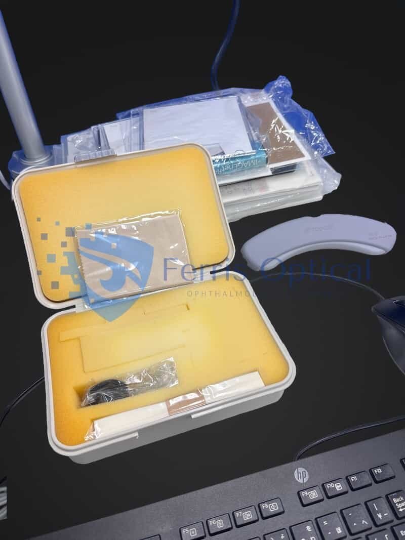
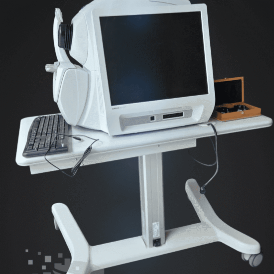
- 2021 Topcon DRI OCT Triton Plus
- PC-Set
- Vorderkammer-Linsen-Einheit AA-1
Referenz
 English
 Spanish
 German
 French