
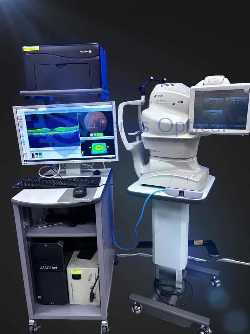
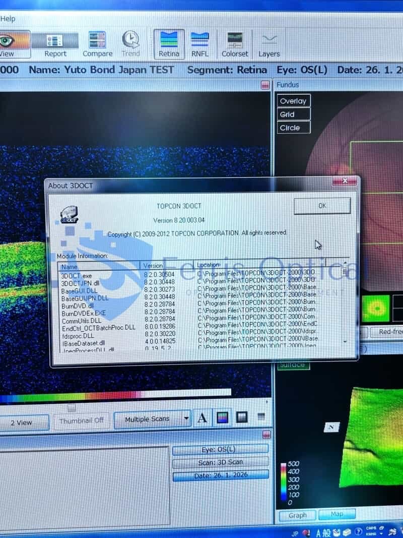
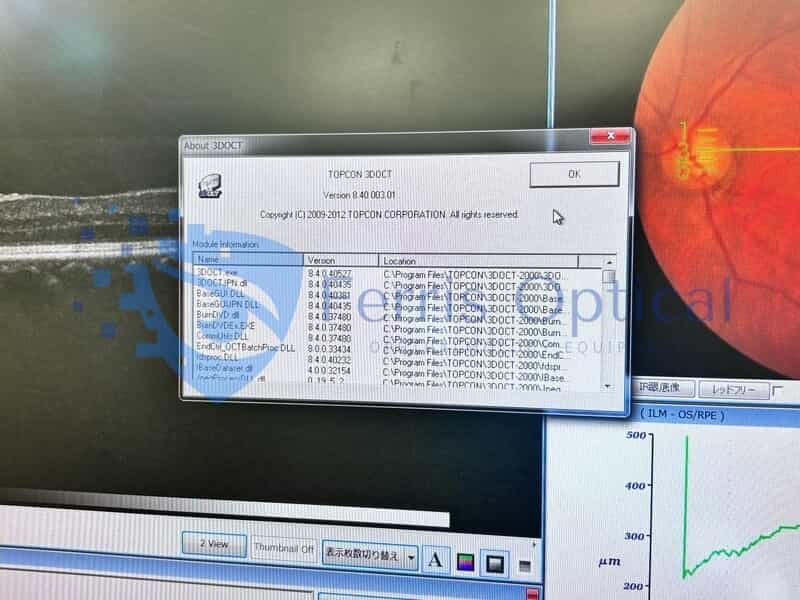
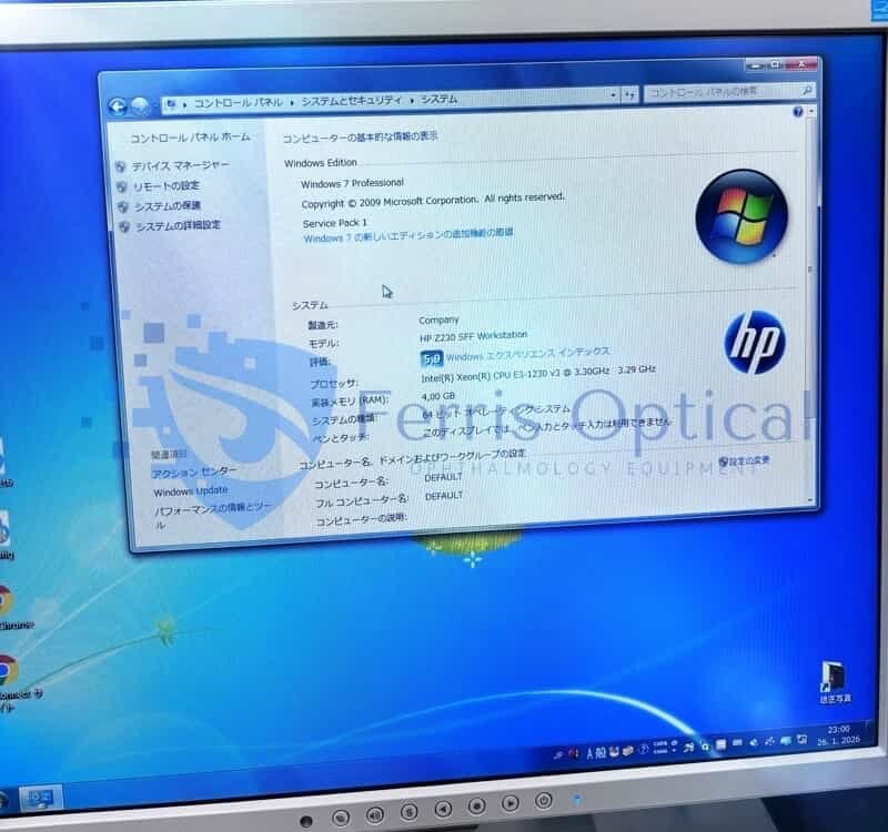
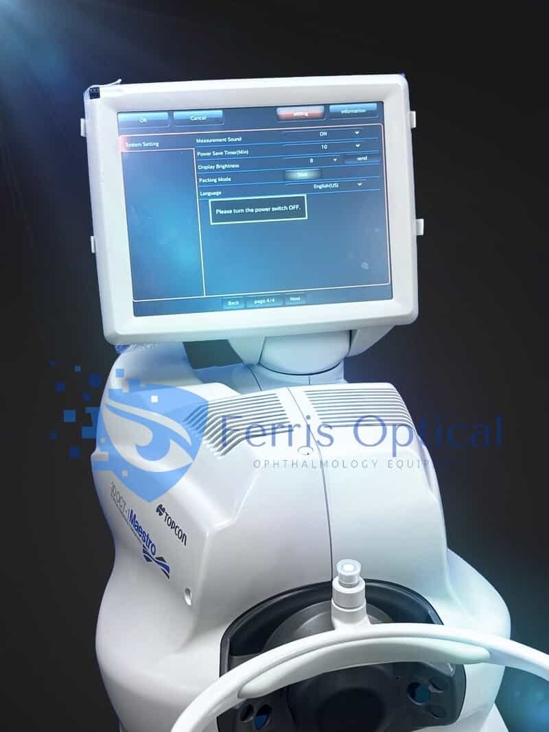
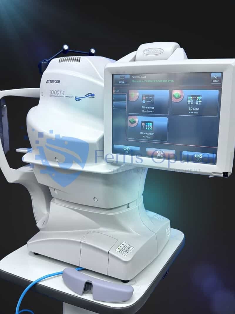
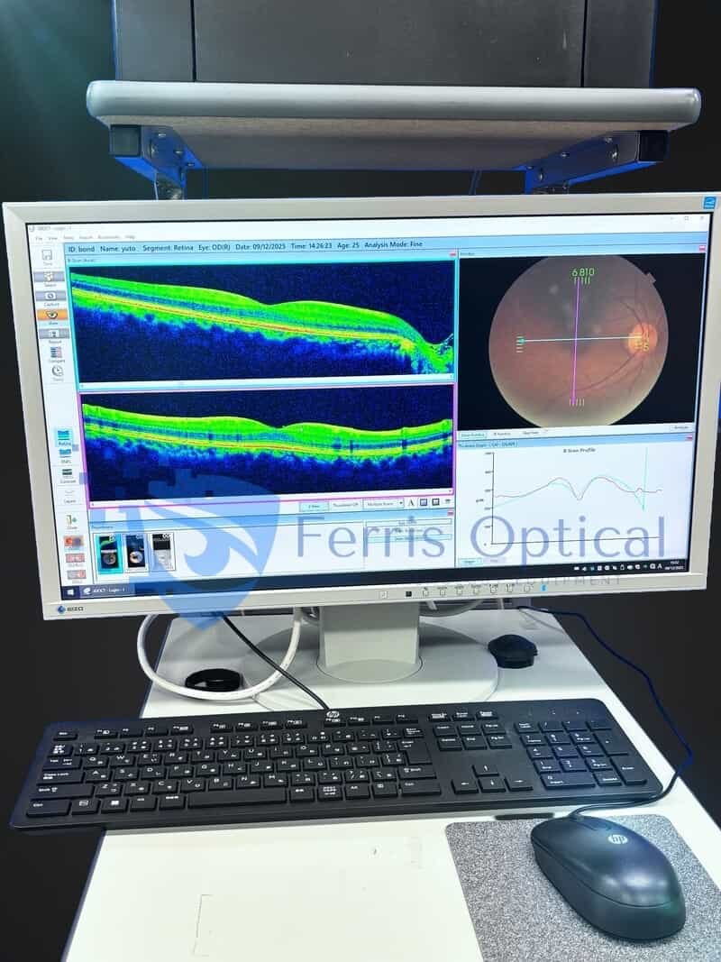
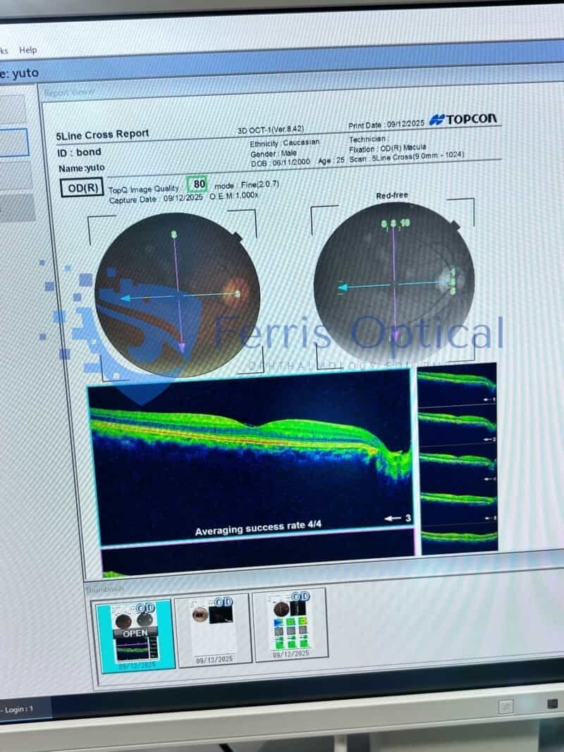
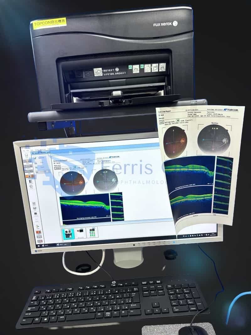
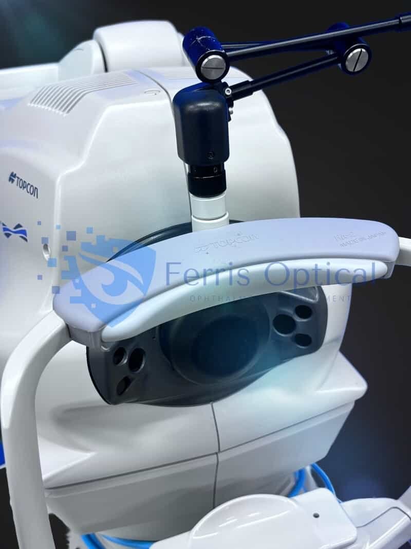
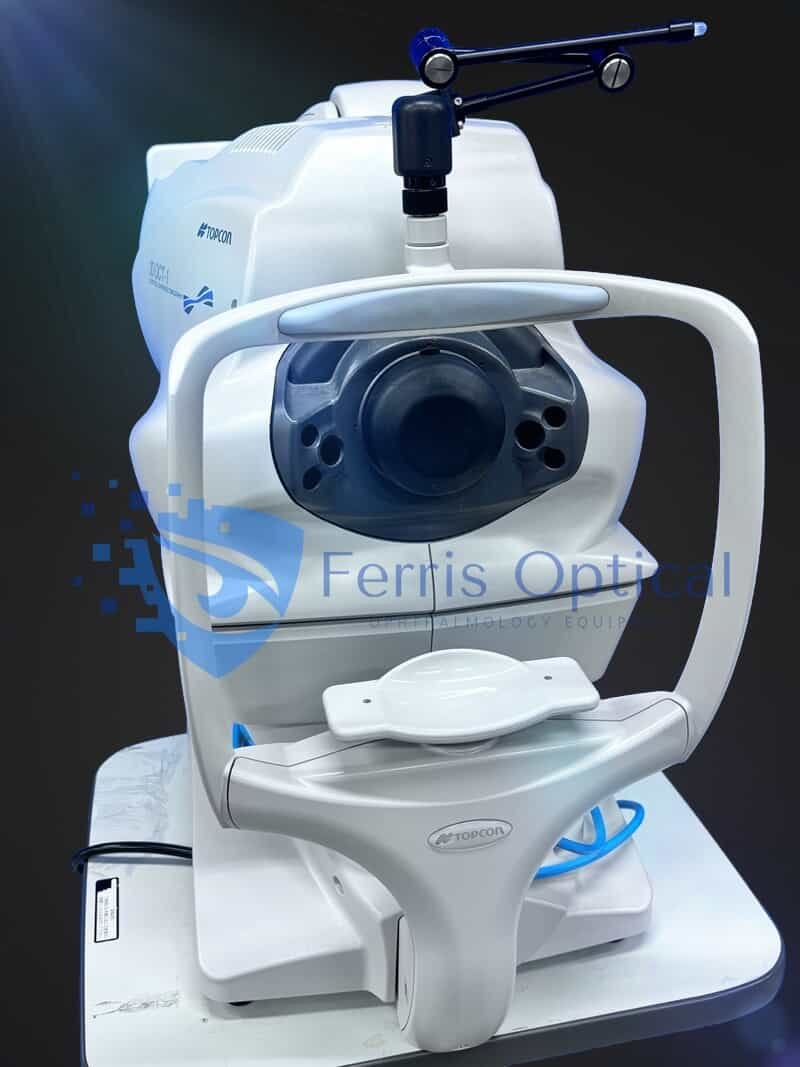
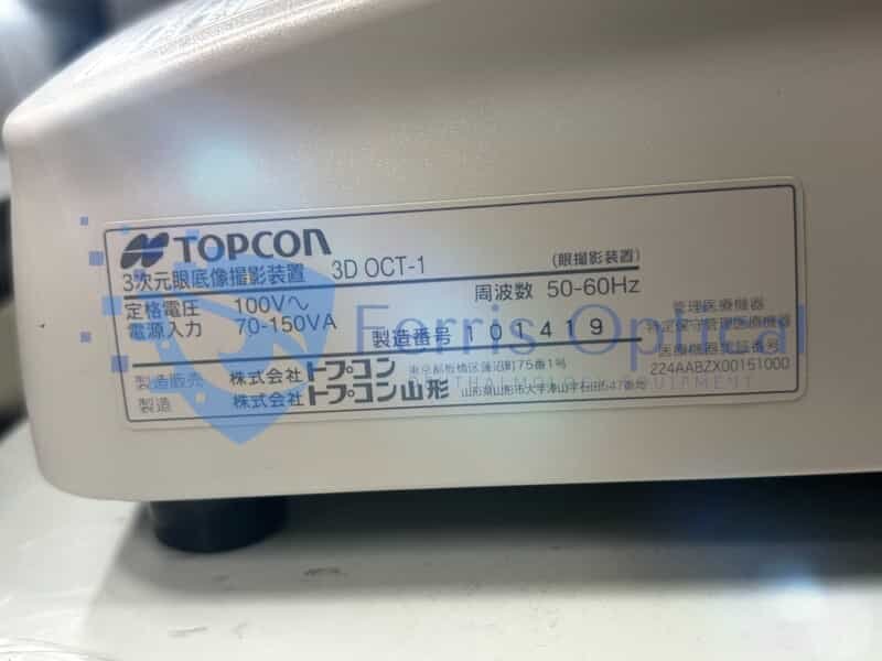
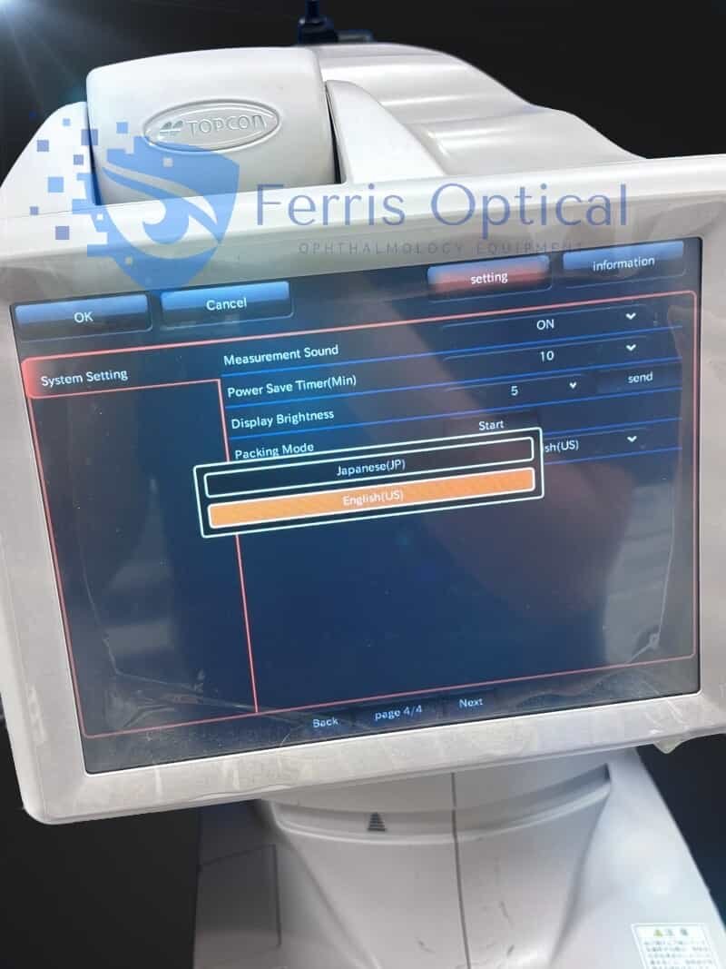
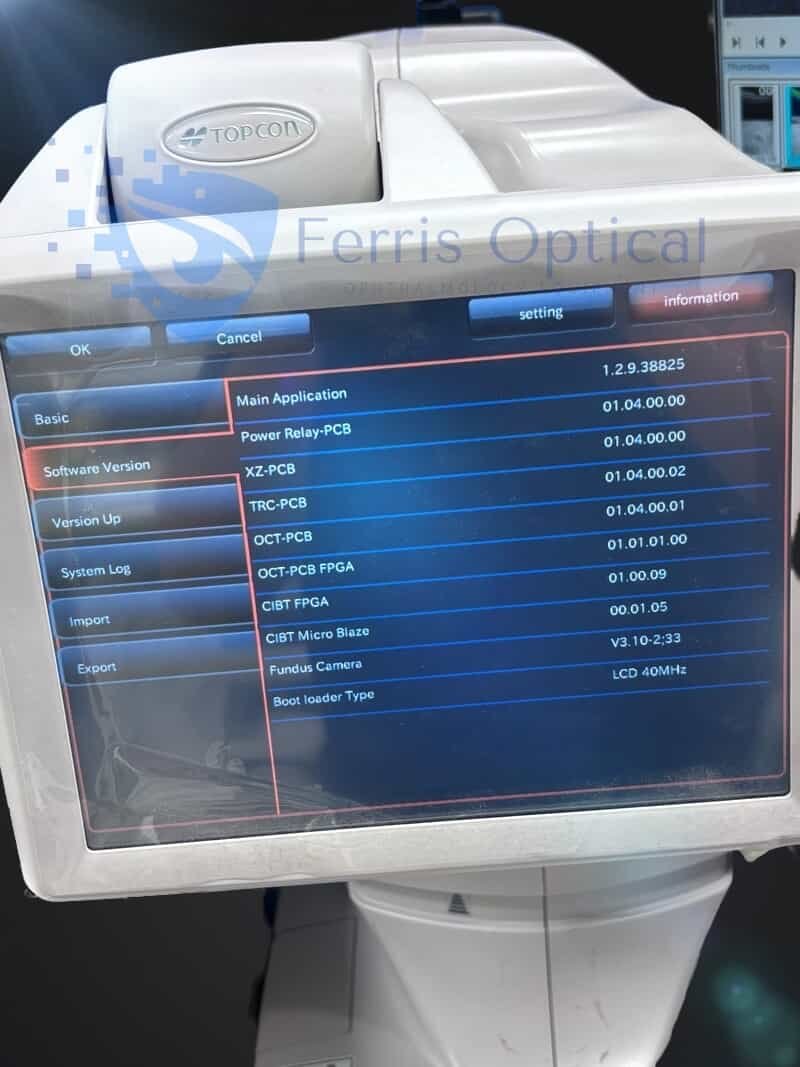
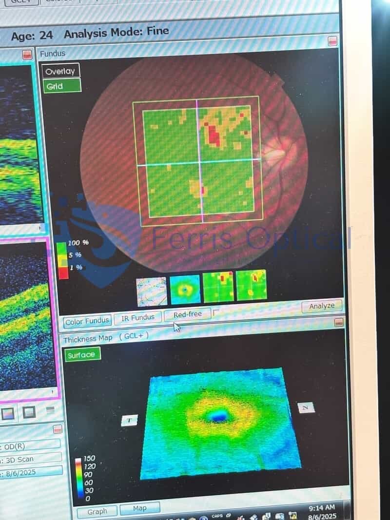
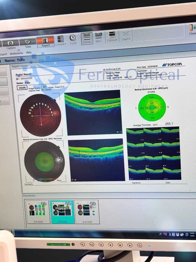
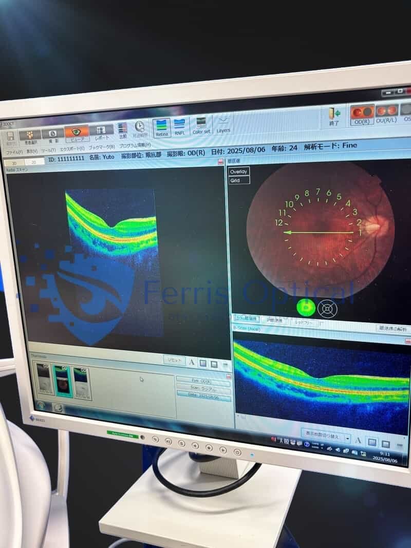
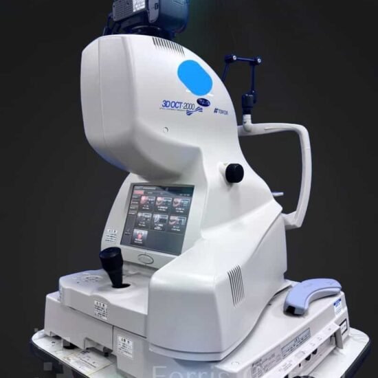
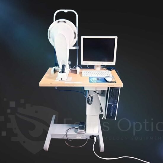
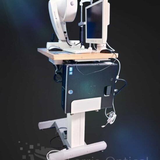
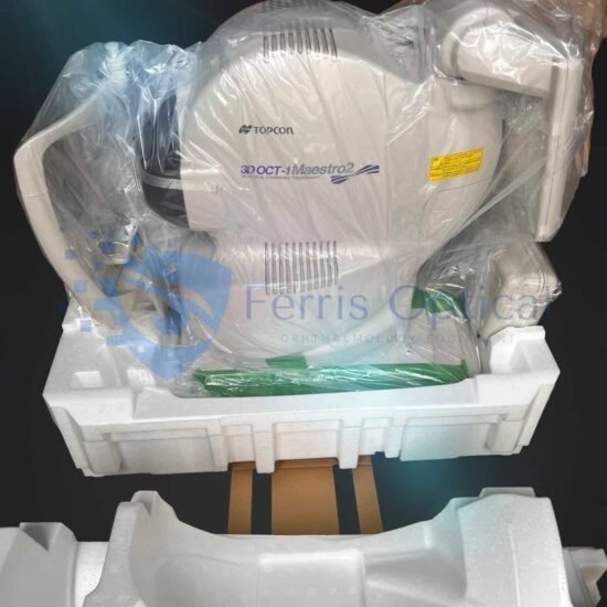
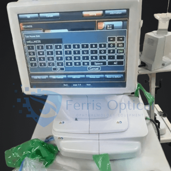
The Topcon Maestro 1 OCT/Fundus Camera is a fully integrated OCT and color fundus imaging system designed for fast, accurate retinal diagnostics. This reconditioned unit delivers reliable performance with automated alignment, high-resolution OCT scans, and color fundus photography in a compact footprint.
The Topcon Maestro 1 combines spectral-domain OCT and color fundus photography into a single, space-efficient platform designed to streamline retinal imaging workflows in busy clinical environments. With its fully automated capture, the Maestro 1 minimizes operator dependency by automatically aligning, focusing, and capturing images with a single touch. This ensures consistent image quality while reducing patient chair time and improving overall efficiency.
TOPCON Maestro 1 Key Features
1) Fundus photography
In addition to the plethora of other imaging modalities on the Topcon Maestro, it can capture single-frame fundus photos simultaneously with other scans. Despite being one of the oldest forms of ophthalmic imaging, fundus photography remains an essential part of today’s clinical practice, from documenting retinal lesions to monitoring glaucomatous optic nerve changes. The Topcon Maestro can also compile montages of several photos taken in different retinal quadrants.
2) Retina/macula OCT
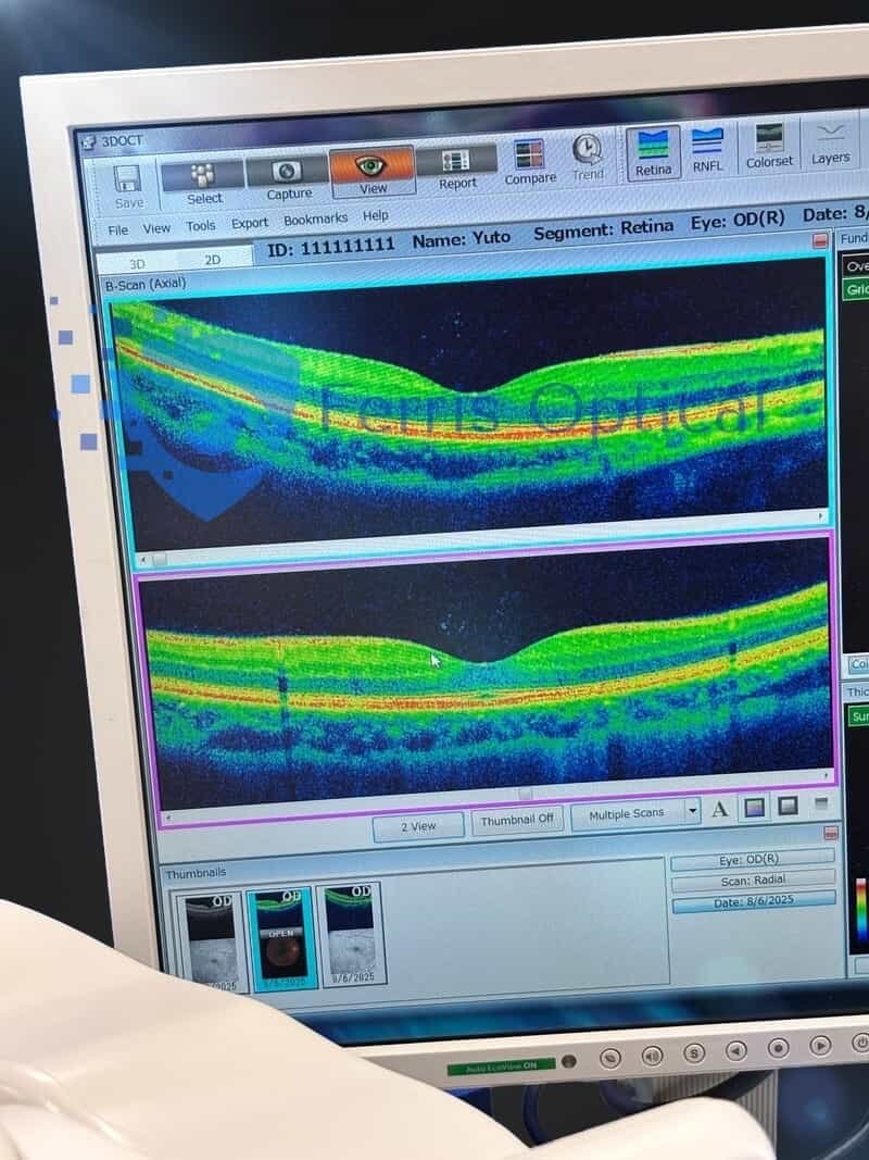
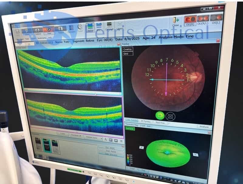
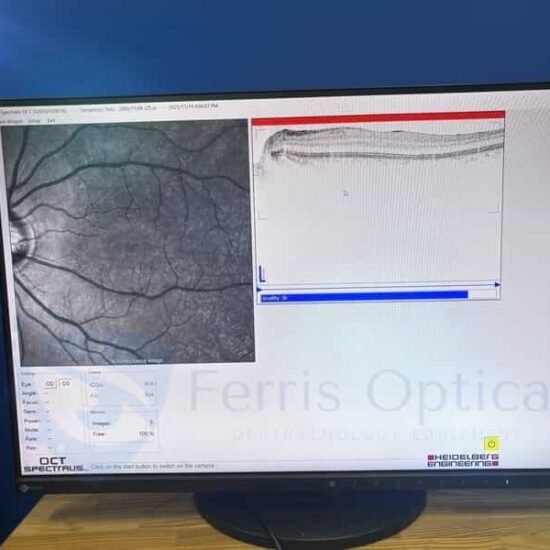
The ability to assess disorders of the retina and macula via OCT is essential for clinical practice today. The Topcon Maestro’s ability to perform single-scan OCT and cube raster scans of the macula enhances the diagnosis and treatment of retinal pathology. All OCT images are taken simultaneously with a corresponding photograph.
3) Optic nerve OCT
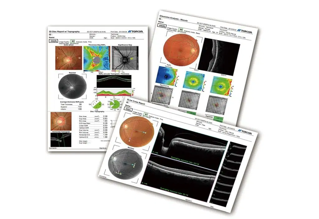
Having a technique to image and assess the optic nerve is crucial for today’s eye care practitioners. OCT has revolutionized the way we diagnose and treat optic nerve pathology. This provides the clinician a simple way to assess optic nerve structure, as well as the integrity of the retinal nerve fiber (RNFL) and ganglion cell layers.
As seen in Figure 6 of the optic nerve OCT scan for primary open-angle glaucoma of the right eye, the red-free filter is also a nice feature, allowing for a closer look at various disorders of the retina and optic nerve. Here, the red-free image shows a superior wedge defect that corresponds with RNFL thinning.
4) Glaucoma: anterior segment (pachymetry and anterior chamber depth)
Gone are the days of invasive techniques to assess corneal thickness. Anterior segment OCT makes pachymetry measurements simple and easy (and is a billable procedure to boot!).
In Figure 8, we see pachymetry taken via the Topcon Maestro’s anterior segment OCT.
5) Assessing scleral contact lenses
Last but not least, we use this instrument’s anterior-segment capabilities to assess the fit of scleral contact lenses. Determining adequate central clearance, as well as satisfactory scleral landing zones, are two ways to implement anterior segment OCT when fitting scleral lenses.
While many other factors and considerations go into the final scleral lens fitting, implementing anterior segment OCT can facilitate and optimize the fitting process for clinicians.
TOPCON Maestro 1 Specifications:
| OBSERVATION & PHOTOGRAPHY OF FUNDUS: | |
| Type of photography | Color, Red-free* & IR*** |
| Picture angle of photography | 45° ± 5% or less |
| 30° or equivalent (digital zoom) | |
| Operating distance | 34.8 ± 0.1mm (when taking a picture of fundus) |
| Photographable diameter of pupil | Normal pupil diameter: ø4.0mm or more |
| Small pupil diameter: ø3.3mm or more | |
| Fundus image resolution (on fundus) | Center: 60 lines/mm or more |
| Middle (r/2) : 40 lines/mm or more | |
| Middle (r) : 25 lines/mm or more | |
| IR photography : Center: 5 lines/mm or more*** | |
| OBSERVATION & PHOTOGRAPHY OF FUNDUS TOMOGRAM: | |
| Scan range (on fundus) Horizontal direction | 3 – 12mm ± 5% or less |
| Vertical direction | 3 – 9mm ± 5% or less |
| Scan pattern | 3D scan (horizontal/vertical) |
| Linear scan (Line-scan/Cross-scan/Radial-scan) | |
| Scan speed | 50,000 A-Scans per second |
| Lateral resolution | 20µm or less |
| In-depth resolution | 6µm or less |
| Photographable diameter of pupil | ø2.5mm or more |
| OBSERVATION & PHOTOGRAPHY OF FUNDUS IMAGE / FUNDUS TOMOGRAM: | |
| Fixation target | Internal fixation target: |
| Dot-matrix type organic EL | |
| The display position can be changed and adjusted. | |
| The displaying method can be changed. | |
| Peripheral fixation target: | |
| This is displayed according to the internal fixation target displayed position. | |
| External fixation target | |
| OBSERVATION & PHOTOGRAPHY OF ANTERIOR SEGMENT: | |
| Type of photography | Color & IR |
| Operating distance | 62.6 ± 0.1mm (when taking a picture of anterior segment)** |
| OBSERVATION & PHOTOGRAPHY OF ANTERIOR SEGMENT TOMOGRAM: | |
| Operating distance | 62.6 ± 0.1mm (when taking a picture of anterior segment)** |
| Scan range (on cornea)** | Horizontal direction: 3 – 6mm ± 5% or less |
| Vertical direction: 3 – 6mm ± 5% or less | |
| Scan pattern | Linear scan (Line-scan/Radial-scan) |
| Scan speed | 50,000 A-Scans per second |
| Fixation target | External fixation target |
* Digital red-free photography that processes a color image and displays it in pseudo-red-free condition.
** Observation & photography of the anterior segment can be performed only when the anterior segment attachment (HA-2) is used.
*** This is used only for recording the position where a tomogram is captured.
Include:
- TOPCON Maestro 1 – 3D OCT-1
- Windows 11 workstation
- Table
- Topcon IMAGEnet 6 software
- Color Fundus Imaging
- Red-Free Imaging
- Fluorescein Angiography (FA)
- Fundus Autofluorescence (FAF)
- One-year Warranty (no additional cost)
- Full-featured All-in-One Computer
- Manual on USB drive
- Professionelles Packen
Referenz
 English
 Spanish
 German
 French