
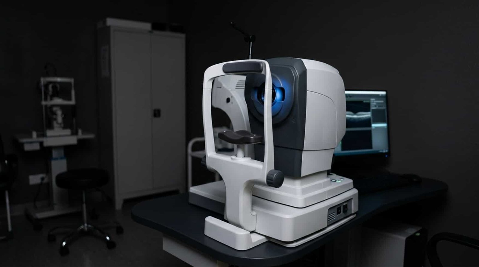
Die aufkommende Technologie, bekannt als optische Kohärenztomographie (OCT), entwickelt sich schnell zu einem der wichtigsten Werkzeuge in der Medizin. Diese Reichweite beschränkt sich nicht nur auf die Medizin, sondern erstreckt sich auch auf die biomedizinische Forschung als Ganzes. Sie wird auch in der pharmazeutischen Entwicklung, bei komplexen Labortests und in der klinischen Praxis eingesetzt. Ursprünglich wurde sie jedoch für die Ophthalmologie entwickelt.
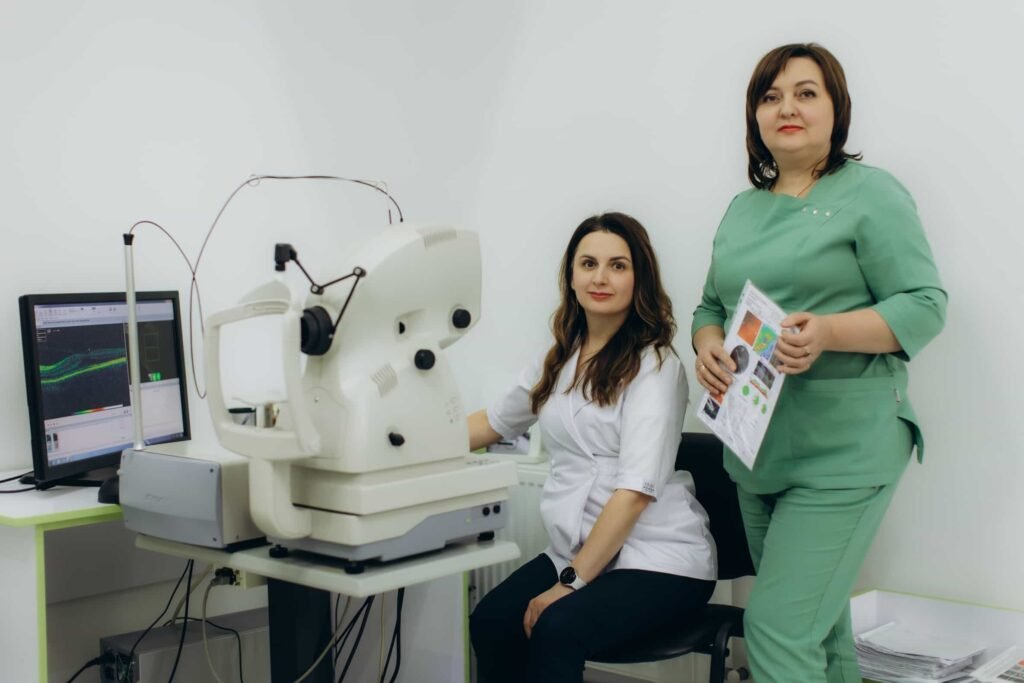
In der Augenheilkunde haben Optometristen und Augenärzte ihre Praxen revolutioniert und die Art und Weise, wie sie Augenkrankheiten, einschließlich Glaukom, Netzhauterkrankungen und Hornhauterkrankungen, diagnostizieren und behandeln, grundlegend verändert. In Labor- und biomedizinischen Umgebungen kann die optische Kohärenztomographie Bildgebungsfähigkeiten für Biomaterialien, die Charakterisierung von Gewebe und die regenerative Medizin bereitstellen. Viele Kliniken nutzen die OCT zur Überwachung des Krankheitsverlaufs oder der Wirksamkeit einer Therapie.
Die Entwicklung der optischen Kohärenz-Tomographie kann die Auswahl eines Systems erschweren, das den Bedürfnissen Ihrer Organisation entspricht. Mehrere Anbieter bieten Lösungen an, die Softwarefunktionen, Forschungsbestandteile, Merkmale, Bildgebungsgeschwindigkeit und Auflösung umfassen. In diesem umfassenden Leitfaden werden wir OCT und seine potenziellen Anwendungen in Ihrer Klinik hervorheben.
OCT-Technologie im Fokus
Die OCT-Bildgebungstechnologie nutzt kohärente Interferometrie, um hochauflösende Querschnittsbilder von Gewebestrukturen zu erstellen. Sie ähnelt dem Ultraschall; anstelle von Schallwellen verwendet sie jedoch Licht. Da die Wellenlänge des Lichts kürzer ist als die von Schall, kann OCT eine Auflösung im Mikrometerbereich erreichen. Dadurch erzielt sie eine tiefere Bildgebung, die eine genauere Untersuchung von Strukturen ermöglicht.
Die optische Kohärenztomographie misst das reflektierte Licht von Gewebeschichten und erstellt detaillierte Bilder von inneren Strukturen. Viele moderne OCT-Systeme können dreidimensionale Volumendatensätze und zweidimensionale Querschnittsbilder erstellen. Die Bilder liefern wichtige Informationen über die strukturelle Integrität, Dicke und Morphologie von Geweben, die über das hinausgehen, was herkömmliche Bildgebung leisten kann.
Einer der Hauptvorteile der OCT ist, dass sie kontaktlos und nicht-invasiv ist. Diese Bildgebung kann schnell durchgeführt werden, ohne Patienten oder Gewebeproben schädlicher ionisierender Strahlung auszusetzen. Ihre Kombination aus Sicherheit, Geschwindigkeit und Neuheit hat viele Praktiker dazu veranlasst, ihre Technologie zu nutzen.
OCT Ophthalmologische Anwendungen
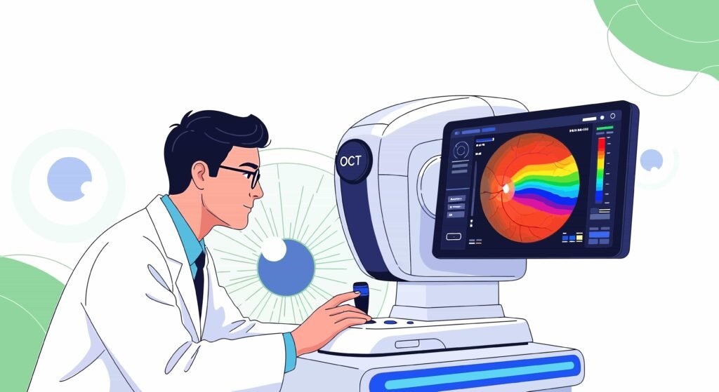
Die OCT-Technologie ist der Goldstandard für Diagnose und Überwachung mehrerer Augenerkrankungen. Bis heute ist die Augenheilkunde die beständigste Anwendung für OCT. Viele Praktiker geben an, dass ihre Praxen ohne sie nicht so reibungslos laufen würden.
Einer der wichtigsten Anwendungsbereiche ist die Netzhautabbildung. Die OCT ermöglicht die Visualisierung der Netzhautschichten und unterstützt die Diagnose von Netzhautvenenverschlüssen, diabetischen Makulaödemen, altersbedingter Makuladegeneration und anderen Netzhauterkrankungen. Die Beurteilung von Behandlungserfolgen und die Verfolgung des Krankheitsverlaufs können mit eindeutiger Genauigkeit erfolgen.
Die Bildgebung mittels optischer Kohärenztomographie ist oft ein wesentlicher Bestandteil der Glaukombehandlung. Durch die Messung der Dicke des Sehnervenkopfes und der Netzhautnervenfaserschicht können Ärzte strukturelle Veränderungen im Zusammenhang mit einem Glaukom erkennen. Diese Messung wird durchgeführt, bevor es zu einem erheblichen Sehverlust kommt.
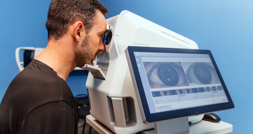
Zusätzlich liefert das anteriore Segment-OCT Bilder der Hornhaut, der Kammerwinkelstrukturen und der Vorderkammer. Die Fähigkeiten unterstützen auch die Beurteilung von Anomalien des vorderen Augenabschnitts, die Planung refraktiver Chirurgie und die Bewertung von Hornhauterkrankungen.
Biomedizinische Forschung
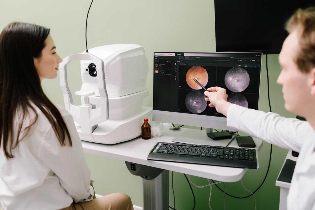
Forscher im Bereich Biomedizin nutzen die OCT zur Untersuchung zellulärer Organisation, Gewebearchitektur und Krankheitsmechanismen. Die OCT ergänzt Mikroskopie und Histologie, indem sie strukturelle Echtzeitinformationen liefert. Forscher, die sich mit Tissue Engineering beschäftigen, nutzen die OCT oft zur Beurteilung der Reifung von konstruiertem Gewebe, zur Überwachung des Zellwachstums und zur Bewertung der Entwicklung von Gerüsten.
In der Neurowissenschaft unterstützt die OCT die Bildgebung von experimentellen Krankheitsmodellen und Nervengewebe. Forscher können strukturelle Veränderungen im Zusammenhang mit Verletzungen und Degeneration untersuchen. Viele erklären, dass diese Spitzentechnologie auch ihr Verständnis der Krankheitsentwicklung verbessert.
Krebsforscher nutzen die OCT zur Beurteilung der Resektionsränder und zur Charakterisierung von Tumoren. Sie ist kein Ersatz für die Pathologie. Sie kann jedoch schnelle strukturelle Informationen liefern, die Untersuchungen und Forschungsarbeiten unterstützen.
Pharmazeutische und biotechnologische Anwendung
Biotechnologie- und Pharmaunternehmen integrieren die optische Kohärenztomographie (OCT) zunehmend in ihre Entwicklungsmodelle, Arbeitsabläufe und Forschungsbereiche. Diese Technologie unterstützt auch die Entwicklung von Biomarkern, Sicherheitsbewertungen, präklinische Studien sowie die Beurteilung der Wirksamkeit von Medikamenten.
OCT bietet eine Längsbildgebung einer Gewebeprobe über die Zeit. Es ermöglicht Forschern, biologische Veränderungen während der Untersuchung zu verfolgen und die Variabilität zu reduzieren. Arzneimittelentwickler können auch strukturelle Reaktionen auf experimentelle Therapien durch Minimierung invasiver Verfahren bewerten.
Biotechnologie-Organisationen konzentrieren sich auf augenheilkundliche Therapien, die oft auf OCT-Endpunkten basieren. OCT-Scans können quantitative Messungen liefern, die als objektive Indikatoren für die Wirksamkeit der Behandlung dienen.
Haupttypen von OCT-Systemen
Bei der Bewertung von OCT-Plattformen sollten Praktiker die primären Technologiekategorien verstehen.
- Die spektrale Domänen-OCT (SD-OCT) wird in der Augenheilkunde weiterhin weit verbreitet und häufig eingesetzt. Diese OCT-Systeme bieten eine hervorragende Bildqualität, schnelle Erfassungsgeschwindigkeiten und starke diagnostische Fähigkeiten.
- SS-OCT (Swept-Source OCT) verwendet einen abstimmbaren Laser. Es bietet eine tiefe Gewebedurchdringung, schnelle Bildgebungsgeschwindigkeiten und eine verbesserte Visualisierung bestimmter Gewebestrukturen. Viele fortgeschrittene augenheilkundliche Kliniken und Forschungszentren beschaffen sich dieses Swept-Source OCT.
- OCT-Systeme für den Forschungsbereich bieten häufig individuell anpassbare Bildgebungsprotokolle, Integrationsmöglichkeiten und Flexibilität. Jede dieser Funktionen ist für akademische Einrichtungen und Forschungsgruppen von großem Wert.
Zu berücksichtigende Faktoren bei der Auswahl eines OCT-Geräts
Primärer Anwendungsfall definiert
Einer der ersten Schritte bei der Auswahl eines OCT-Systems besteht darin, festzulegen, wie es in Ihrer Einrichtung eingesetzt werden soll. Klinische Augenarztpraxen haben andere Anforderungen als Pharmaunternehmen oder Forschungslabore.
Ein auf Netzhauterkrankungen spezialisierter Kliniker könnte Workflow, spezifische Analysen für Krankheiten oder Bildqualität priorisieren. Forschungslabore benötigen möglicherweise anpassbare Protokolle, erweiterte Analysefähigkeiten oder exportierbare Datensätze. Wenn Sie Ihr Hauptziel verstehen, können Sie die verfügbaren Optionen eingrenzen.
Bildauflösungsbewertung
Die Bildauflösung beeinflusst direkt die Fähigkeit, feine strukturelle Details zu visualisieren. Systeme mit höherer Auflösung können subtile Gewebemerkmale enthüllen, die für diagnostische oder Forschungszwecke wichtig sind.
Die axiale und laterale Auflösung sollten beide berücksichtigt werden. Forscher, die mikrostrukturelle Veränderungen untersuchen, profitieren oft von der höchstmöglichen Auflösung, während routinemäßige klinische Praktiken möglicherweise ein Gleichgewicht zwischen Auflösung und Workflow-Effizienz bevorzugen.
Bewertung der Bildgebungsgeschwindigkeit
Moderne OCT-Systeme unterscheiden sich in der Erfassungsgeschwindigkeit. Schnellere Bildgebung verbessert den Patientenkomfort und reduziert Auswirkungen. Hochgeschwindigkeitssysteme können auch den Durchsatz in geschäftigen Labors, Kliniken und Forschungsumgebungen erhöhen.
Organisationen, die große Studien oder klinische Abläufe mit hohem Volumen durchführen, sollten die Gesamtleistung des Arbeitsablaufs und die Scanraten sorgfältig prüfen.
Berücksichtigung der Eindringtiefe
Viele Anwendungen erfordern tiefere Bildgebungskapazitäten. Swept-Source-OCT-Systeme bieten oft eine verbesserte Penetration durch stark streuende Gewebe und können die Visualisierung tieferer anatomischer Strukturen verbessern.
Forschungsgruppen, die mit komplexen Gewebemodellen arbeiten, sollten prüfen, ob eine tiefere Penetration für ihre spezifischen Projekte sinnvolle Vorteile bietet.
Software- und Analytiküberprüfung
Die Fähigkeiten der Software können den Wert eines OCT-Systems erheblich beeinflussen. Fortgeschrittene Softwarepakete können quantitative Messungen, Fortschrittsanalysen und Berichtsfunktionen umfassen.
Klinische Anwender profitieren oft von krankheitsspezifischen Analysemodulen. Forscher legen möglicherweise Wert auf Datenexportfunktionen und den Zugriff auf rohe Bilddatensätze.
Integration und Datenmanagement
Die Integration mit elektronischen Patientenakten, Laborinformationssystemen und Forschungsdatenbanken sollte nicht außer Acht gelassen werden. Effizientes Datenmanagement verbessert den Arbeitsablauf, unterstützt die Einhaltung von Vorschriften und erleichtert die Zusammenarbeit.
Organisationen, die multizentrische Studien durchführen, benötigen möglicherweise standardisierte Datenformate und zentralisierte Bildverwaltungslösungen.
Anbietersupport und Schulung
Der Kauf eines OCT-Geräts stellt eine bedeutende Investition dar. Anbieterunterstützung, Schulungsprogramme und Serviceverträge können den langfristigen Erfolg maßgeblich beeinflussen.
Potenzielle Käufer sollten Reaktionszeiten, Verfügbarkeit des technischen Supports, Richtlinien für Software-Updates und Schulungsressourcen bewerten. Zuverlässiger Support ist besonders wichtig für Organisationen mit begrenzter eigener Bildgebungsexpertise.
Regulatorische und Compliance-Aspekte
Klinische Organisationen sollten sicherstellen, dass ausgewählte Systeme die geltenden regulatorischen Anforderungen in ihrer Region erfüllen. Forschungsgruppen, die regulierte Studien durchführen, sollten ebenfalls die Einhaltung relevanter Standards und Dokumentationsanforderungen überprüfen.
Budget und Gesamtkosten (Total Cost of Ownership)
Der Anschaffungspreis stellt nur einen Teil der Investition dar. Organisationen sollten Wartungskosten, Serviceverträge, Softwarelizenzgebühren, Zubehör und Upgrade-Ausgaben berücksichtigen.
Ein preisgünstigeres System kann sich letztendlich als teurer erweisen, wenn ihm wesentliche Funktionen fehlen oder häufige Upgrades erforderlich sind. Umgekehrt kann der Kauf einer übermäßig ausgefeilten Plattform zu unnötigen Ausgaben führen.
Lieferantenfragen
Käufer sollten den Verkäufern mehrere Schlüsselfragen stellen:
- Wie oft veröffentlichen Sie Software-Updates?
- Welche Analysesoftware ist in der Technologie enthalten?
- Können die gesammelten Rohdaten exportiert werden?
- Sind Referenzen von anderen Organisationen verfügbar?
- Welche Schulungsressourcen werden bereitgestellt?
- Für welche Anwendungen kann dieses System optimiert werden?
- Welche Wartungs- und Serviceoptionen stehen zur Verfügung?
Best Practices für eine erfolgreiche OCT-Implementierung
Die erfolgreiche Implementierung von OCT geht über die bloße Auswahl der richtigen Hardware hinaus. Organisationen sollten standardisierte Bildgebungs- und Qualitätssicherungsprotokolle sowie Schulungsprogramme für das Personal einrichten.
Klinische Praxen müssen sicherstellen, dass Techniker eine angemessene Schulung erhalten, um die Konsistenz und Bildqualität zu maximieren. Forschungsorganisationen sollten Standardverfahren für die Bildgebung entwickeln, um die Reproduzierbarkeit zwischen Studien zu verbessern.
Regelmäßige Kalibrierung und Wartung tragen zur Erhaltung der Bildgebungsleistung bei. Die Etablierung klarer Arbeitsabläufe für Datenspeicherung, -analyse und -sicherung erhöht den langfristigen Wert zusätzlich.
Schlussfolgerung
Die optische Kohärenztomographie hat die Bildgebung in Biotechnologie, Ophthalmologie, biomedizinischer Forschung, klinischer Untersuchung und pharmazeutischer Entwicklung verändert und revolutioniert. Ihre Fähigkeit, Gewebestrukturen hochauflösend und nicht-invasiv darzustellen, macht sie zu einem unverzichtbaren Werkzeug für das moderne Gesundheitswesen und wissenschaftliche Entdeckungen.
Die Auswahl der besten OCT-Maschine erfordert eine sorgfältige Bewertung der organisatorischen Ziele, der Bildgebungsanforderungen, der Workflow-Bedürfnisse, der Softwarefunktionen und der Budgetüberlegungen. Kliniken legen möglicherweise Wert auf diagnostische Effizienz und krankheitsspezifische Analysen, während Forschungseinrichtungen oft Flexibilität, Anpassungsmöglichkeiten und erweiterten Datenzugriff benötigen. Pharma- und Biotech-Organisationen konzentrieren sich möglicherweise auf die Fähigkeit zur Längsbildgebung und die Entwicklung von Biomarkern.
Durch die sorgfältige Bewertung dieser Faktoren und die Partnerschaft mit einem zuverlässigen Anbieter können Organisationen eine OCT-Plattform auswählen, die aktuelle Ziele unterstützt und gleichzeitig an zukünftige Anforderungen anpassbar bleibt. Das richtige OCT-System kann die diagnostische Sicherheit verbessern und erhöhen, den Forschungsfortschritt beschleunigen, die Studienqualität verbessern und letztendlich zu besseren wissenschaftlichen und klinischen Ergebnissen beitragen. (Huang et al., 1991; Drexler & Fujimoto, 2015).
Referenzen
Bouma, B. E., & Tearney, G. J. (Hrsg.). (2001). Handbook of optical coherence tomography. Marcel Dekker.
Drexler, W., & Fujimoto, J. G. (2015). Optical coherence tomography: Technology and applications (2. Aufl.). Springer.
Fercher, A. F., Hitzenberger, C. K., Kamp, G. & El-Zaiat, S. Y. (1995). Messung intraokularer Abstände durch spektrale Rückstreuinterferometrie. Optics Communications, 117(1–2), 43–48.
Fujimoto, J. G., Pitris, C., Boppart, S. A., & Brezinski, M. E. (2000). Optische Kohärenztomographie: Eine aufstrebende Technologie für biomedizinische Bildgebung und optische Biopsie. Neoplasia, 2(1–2), 9–25.
Huang, D., Swanson, E. A., Lin, C. P., et al. (1991). Optische Kohärenztomographie. Science, 254(5035), 1178–1181.
Leung, C. K. S., Ye, C., & Weinreb, R. N. Optische Kohärenztomographie im Glaukommanagement.
National Eye Institute. (o. D.). Überblick über die optische Kohärenztomographie (OCT).
Spaide, R. F., Fujimoto, J. G., & Waheed, N. K. (2018). Swept-Source-OCT in der Augenheilkunde. Retina.
Swanson, E. A., Izatt, J. A., Hee, M. R., u. a. (1993). In vivo retinal imaging by optical coherence tomography. Optics Letters.
American Academy of Ophthalmology. (o. D.). Optische Kohärenz-Tomographie Klinische Leitlinien.
 English
 Spanish
 German
 French