
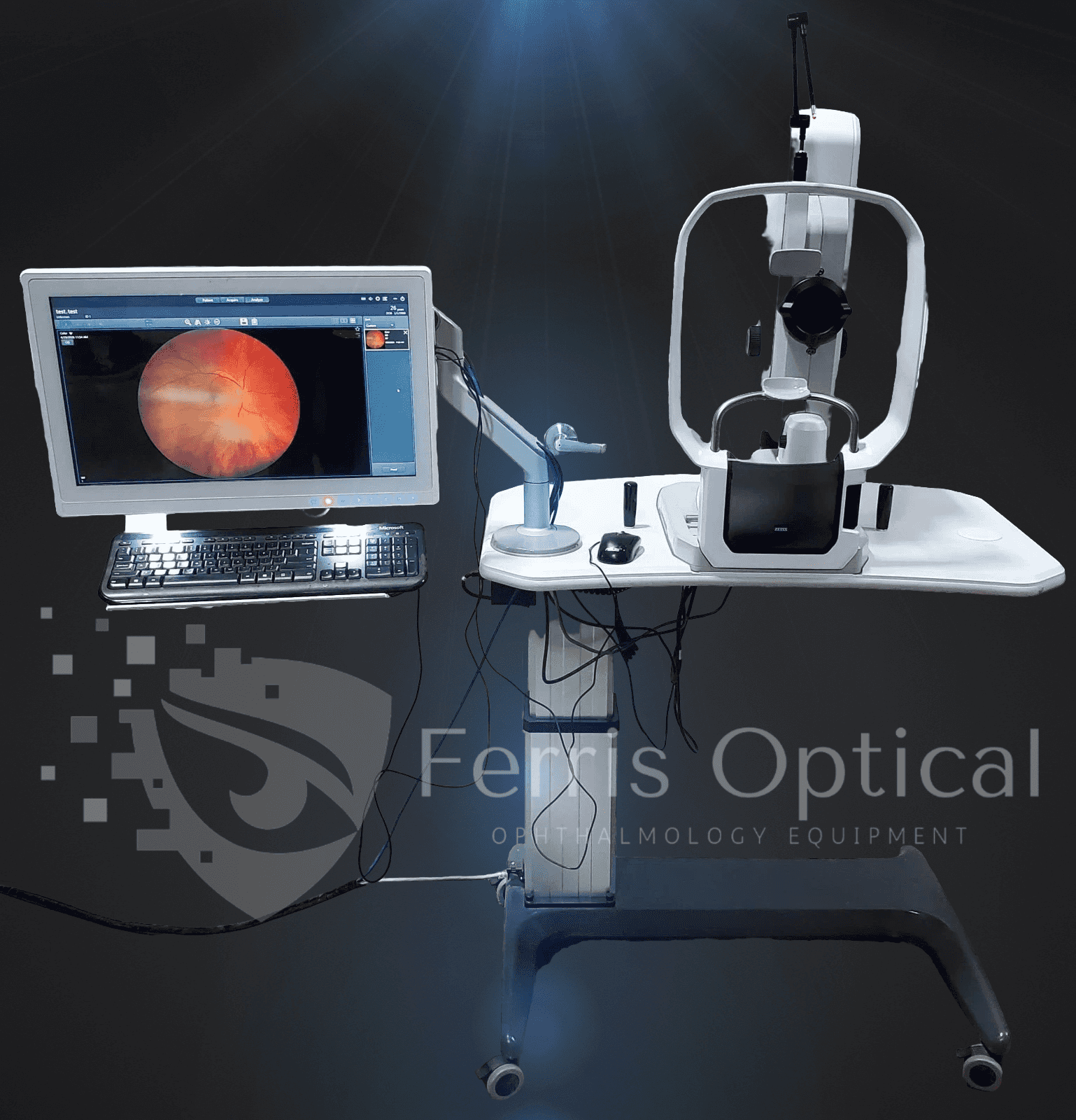
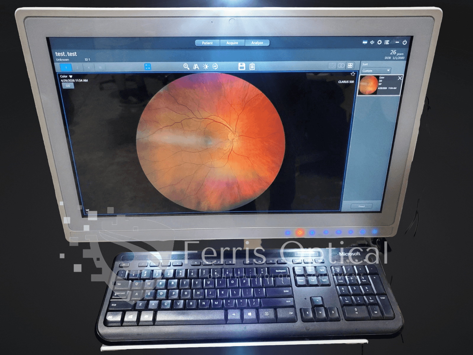
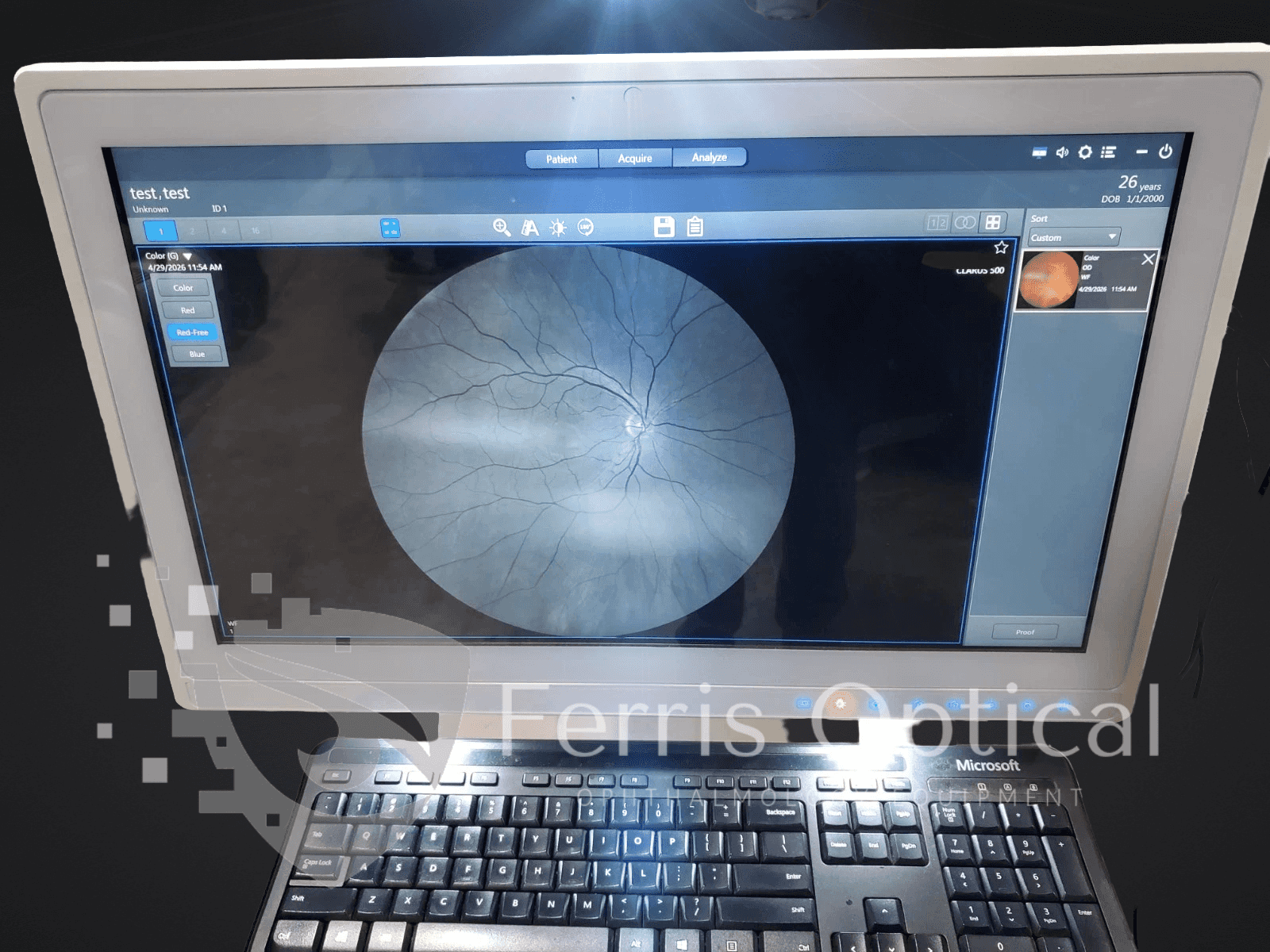
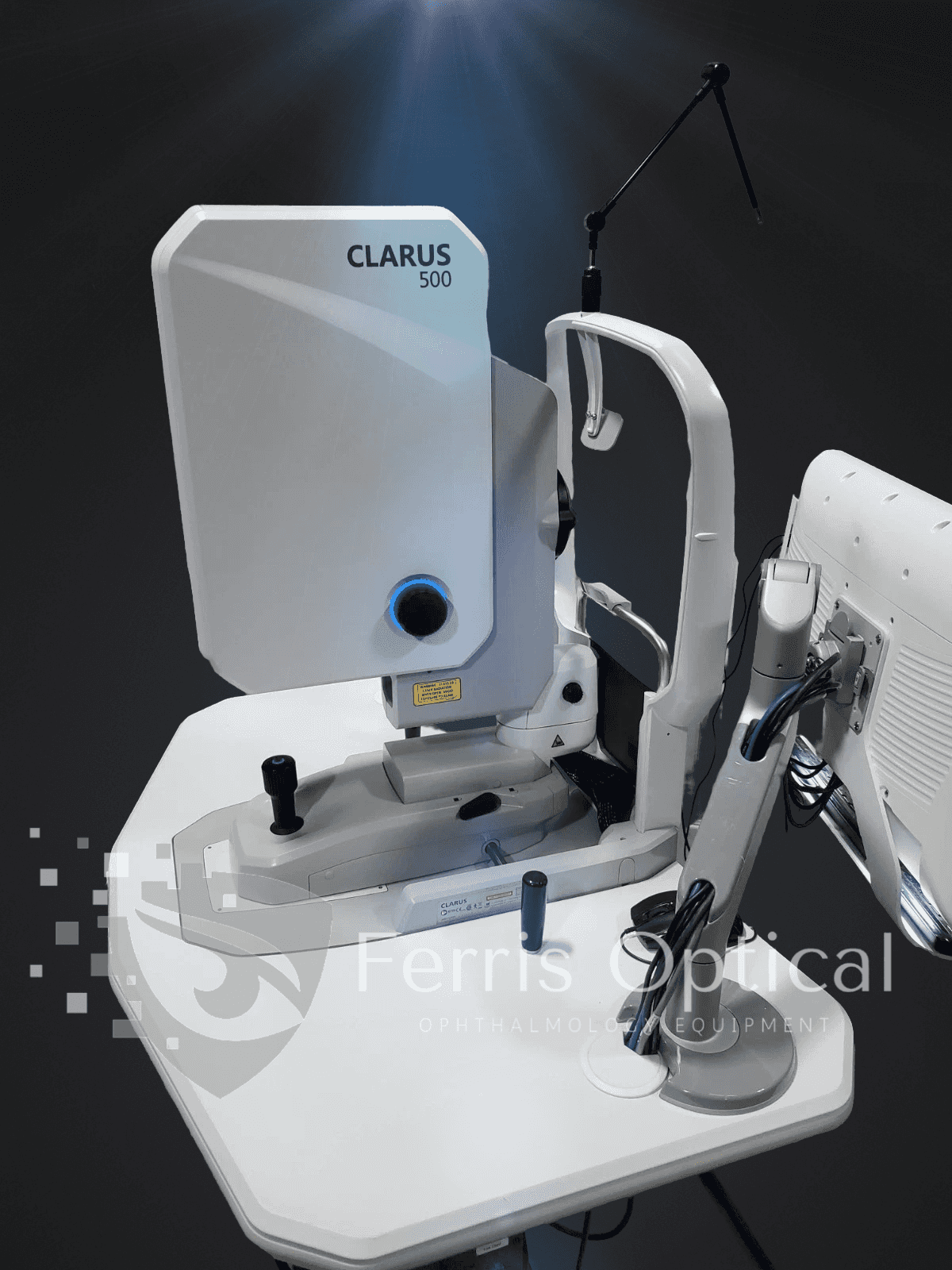
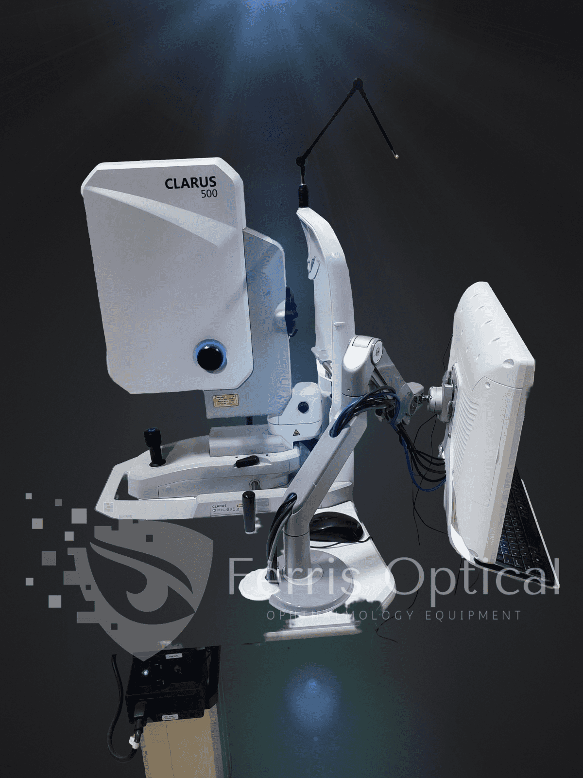
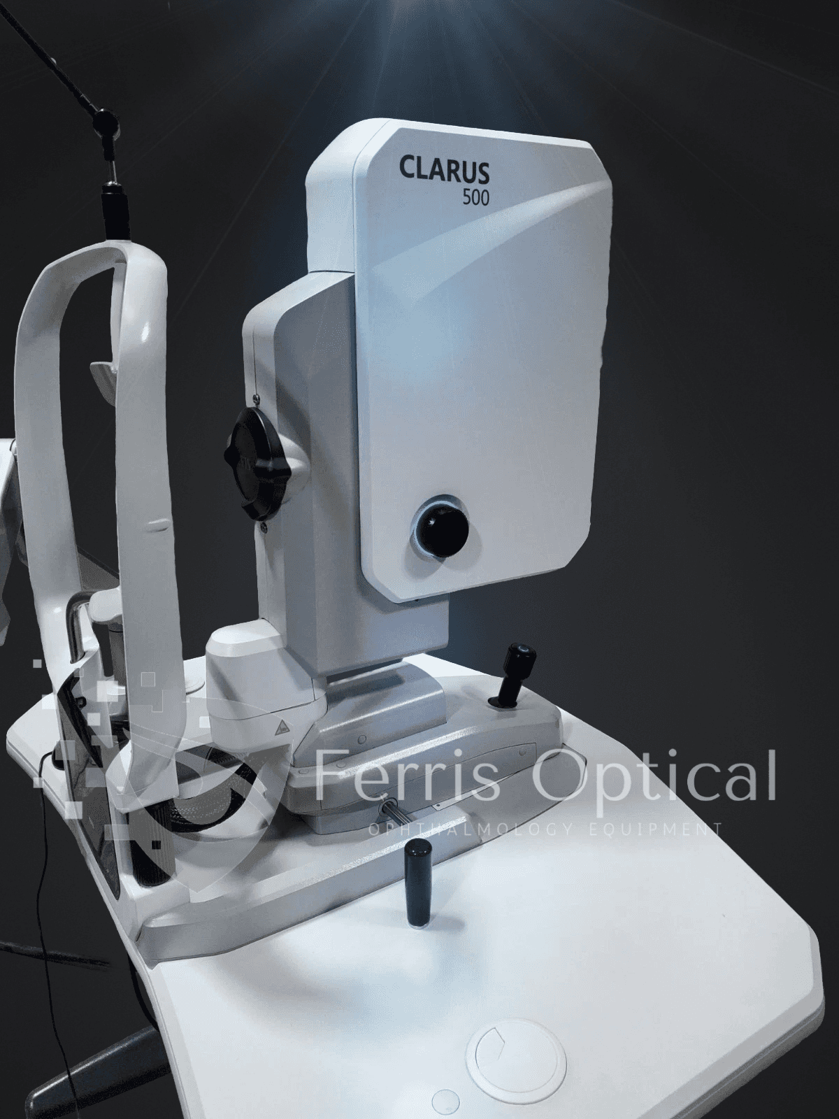
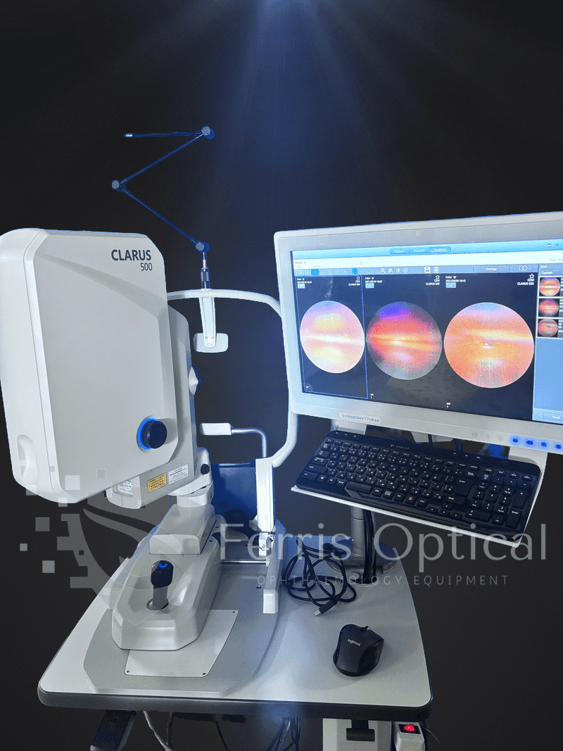
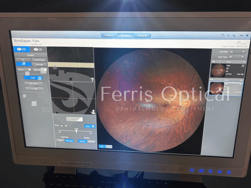
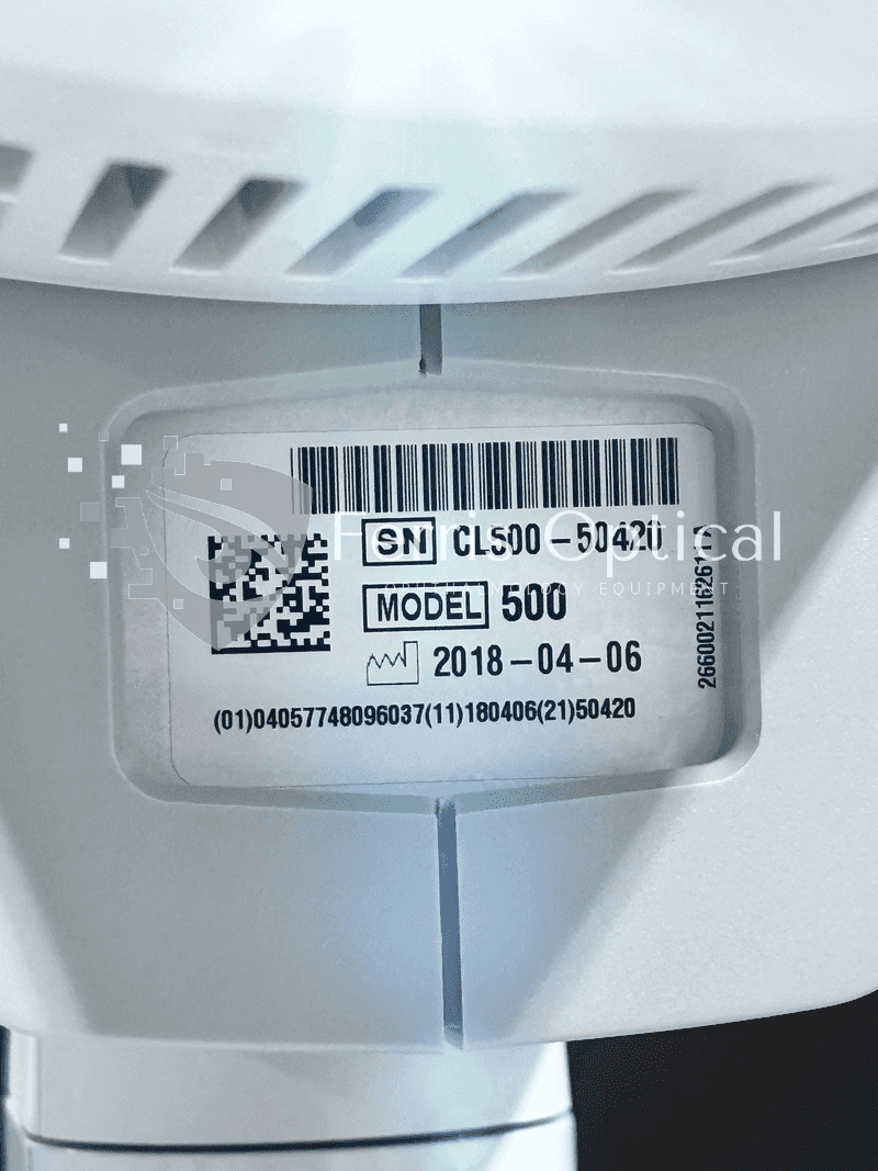
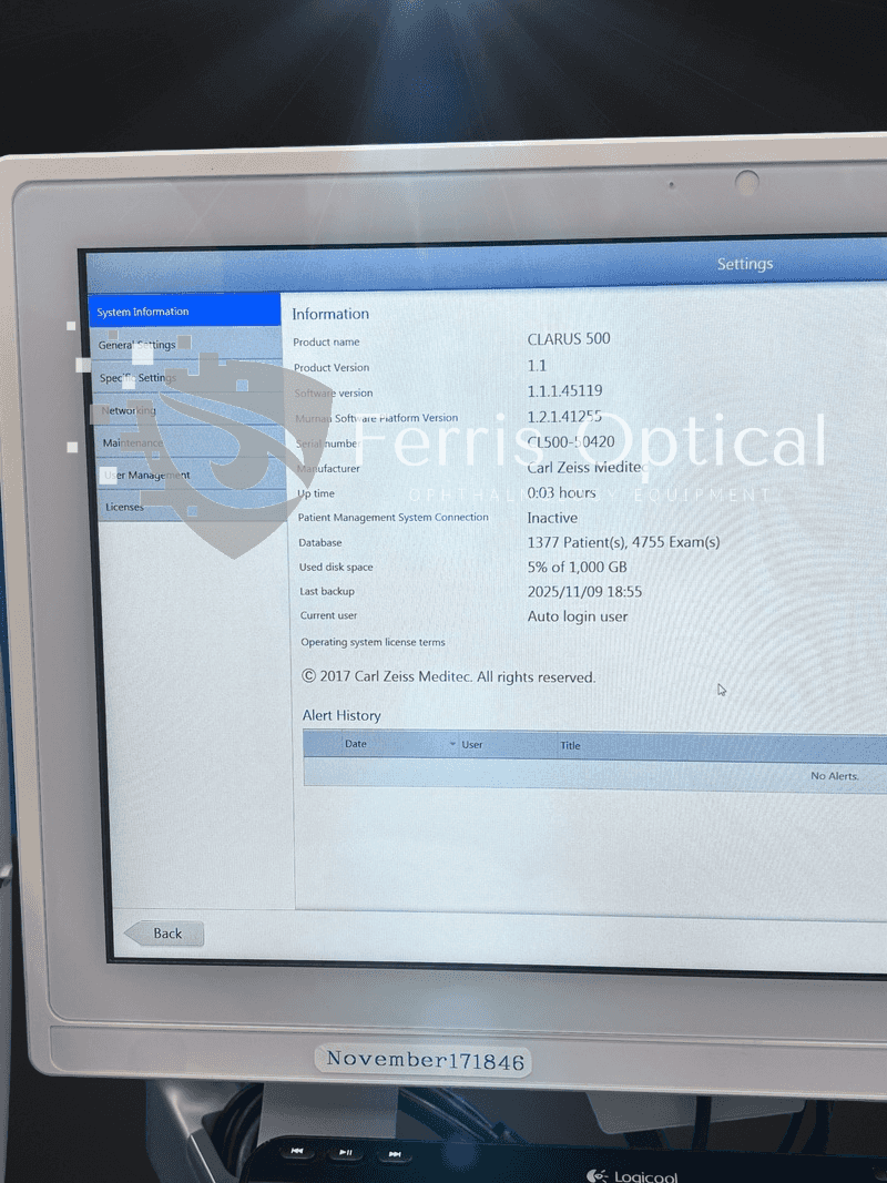
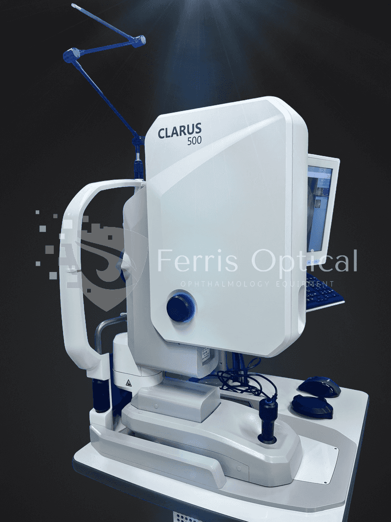
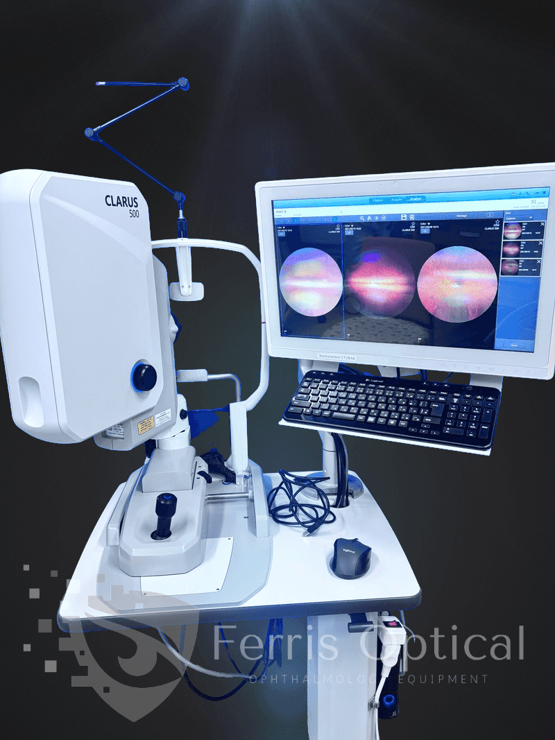
ZEISS CLARUS 500 empowers optometrists to track subtle retinal changes over time with confidence. In addition to producing true-color images, it captures high-resolution fundus autofluorescence (FAF-Blue and FAF-Green) and external eye images. The intuitive review software and ultra-high-resolution imaging help optometrists detect, document, and manage retinal disease progression, leading to better patient outcomes.
Designed for high-volume optometry practices, the CLARUS 500 streamlines diagnostic workflow, enhances patient education, and makes follow-up comparisons easy with consistently high-quality images. Upgrade your clinic’s imaging capabilities—contact us for availability, condition details, included accessories, software options, and shipping.
Unlike older systems, the CLARUS 500 is the first ultra-widefield fundus camera to deliver true color and crystal-clear detail across the entire retina. Optometrists can capture high-resolution images from the macula to the far periphery—all in a single device—streamlining diagnostics and enhancing clinical decision-making.
The Refurbished Zeiss Clarus 500 Ultra-Widefield Camera is an ideal diagnostic tool for optometry clinics seeking state-of-the-art retinal imaging at a practical price. This advanced system offers true-color, high-resolution fundus imaging for precise disease detection, while the fully refurbished unit is tested, calibrated, and certified for reliable performance—helping optometrists deliver excellent patient care and maximize their clinic’s investment.
Key Features & Benefits:
- ✅ Ultra-Widefield Imaging (up to 200°) – Optometrists can capture more retinal detail in a single image, supporting thorough and efficient patient exams.
- ✅ True Color, Red-Free & Autofluorescence Imaging – Visualize retinal pathology with unmatched clarity, supporting early and accurate diagnosis in your optometry clinic.
- ✅ High-Resolution (7.3μm per pixel) – Achieve superior image quality for early detection and monitoring of retinal diseases in optometric practice.
- ✅ Fast & Comfortable for Patients – Non-mydriatic imaging streamlines workflow, reduces patient discomfort, and keeps your clinic running smoothly.
- ✅ Advanced Disease Detection – Ideal for optometrists monitoring diabetic retinopathy, AMD, glaucoma, and other retinal conditions.
- ✅ DICOM Compatible – Seamlessly integrates with EMR systems in optometry clinics for easy and secure data management.
Zeiss Clarus 500 FA — Refurbished Ultra-Widefield Fundus Camera for Optometrists with Fluorescein Angiography
The Zeiss Clarus 500 FA is designed for optometrists who want comprehensive retinal imaging and angiography without compromising workflow or patient comfort. It delivers 133° single-capture ultra-widefield true color fundus photography and fluorescein angiography in a non-contact, non-mydriatic system. Broadband LED illumination provides natural tissue color for accurate assessment, while the refurbished Clarus 500 FA is fully tested, calibrated, and ready for clinical use—offering advanced functionality at a fraction of the cost of new models.
A National Library of Medicine study published in JAMA Ophthalmology confirms that ultra-widefield fluorescein angiography identifies peripheral retinal nonperfusion associated with proliferative diabetic retinopathy — pathology that standard 45°–60° cameras routinely miss.
Why Optometry Clinics Choose the Zeiss Clarus 500 FA
- 133° single-capture ultra-widefield imaging — covers the posterior pole and peripheral retina in one shot; automated montage extends coverage to approximately 200°
- True color broadband LED imaging — natural tissue color from a non-laser illumination source; no red-free scanning laser artifacts that misrepresent subtle lesion color
- Fluorescein angiography (FA) — widefield dynamic angiography for retinal vascular disease, leakage mapping, neovascularization detection, and ischemia staging across the full peripheral retina
- Non-mydriatic for most patients — captures through a 3.3 mm pupil undilated; faster clinic workflow and improved patient comfort compared to dilated fundus photography
- Non-contact imaging — no lens, no applanation; comfortable for patients and fast to reposition between eyes
- Zeiss FORUM integration — longitudinal image comparison and multi-modal overlay within the Zeiss ecosystem; DICOM export for EMR integration
- Compact footprint — smaller physical footprint than scanning laser ultra-widefield systems; fits standard exam lanes and imaging rooms
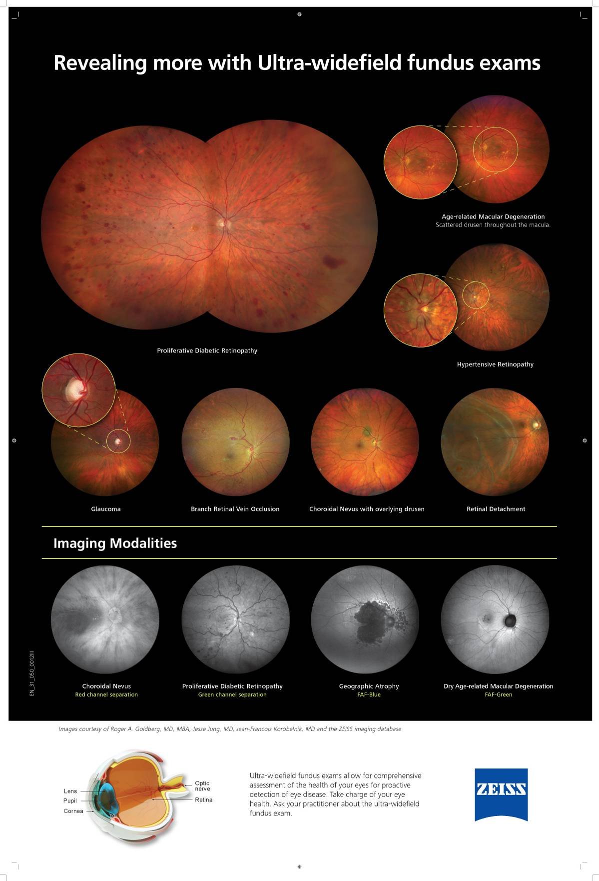
Clinical Applications for Optometrists
The Zeiss Clarus 500 FA supports the full range of posterior segment imaging and angiography workflows in both clinic and imaging center settings.
- Diabetic retinopathy screening — 133° single-capture reveals peripheral neovascularization and ischemia beyond the ETDRS 7-field standard; FA identifies active leakage and neovascular extent
- Retinal vascular occlusion — FA maps ischemic area, collateral development, and macular edema extent across the full posterior pole and periphery
- Peripheral retinal pathology — lattice degeneration, peripheral tears, and detachment extent documented in a single widefield capture for laser treatment planning
- AMD monitoring — posterior pole and mid-peripheral drusen distribution documented; FA identifies active CNV and geographic atrophy boundaries
- Uveitis — widefield FA characterizes peripheral vasculitis and periphlebitis without lens contact in inflamed eyes
- Teleophthalmology and remote screening — 133° non-mydriatic images suitable for remote diabetic retinopathy grading without patient dilation
Who Benefits in Optometry
- Retinal specialists and retina practices — FA plus 133° widefield coverage in one unit for diabetic retinopathy staging, vascular occlusion workup, and AMD management
- Comprehensive ophthalmology clinics — non-mydriatic widefield imaging improves DR screening throughput; FA module adds angiography without a separate dedicated angiographer
- Teleophthalmology and diabetic screening programs — 133° non-mydriatic captures deliver enough peripheral coverage for remote grading at scale
- Practices upgrading from standard 45° cameras — add peripheral coverage and dynamic FA without moving to a scanning laser platform
Includes:
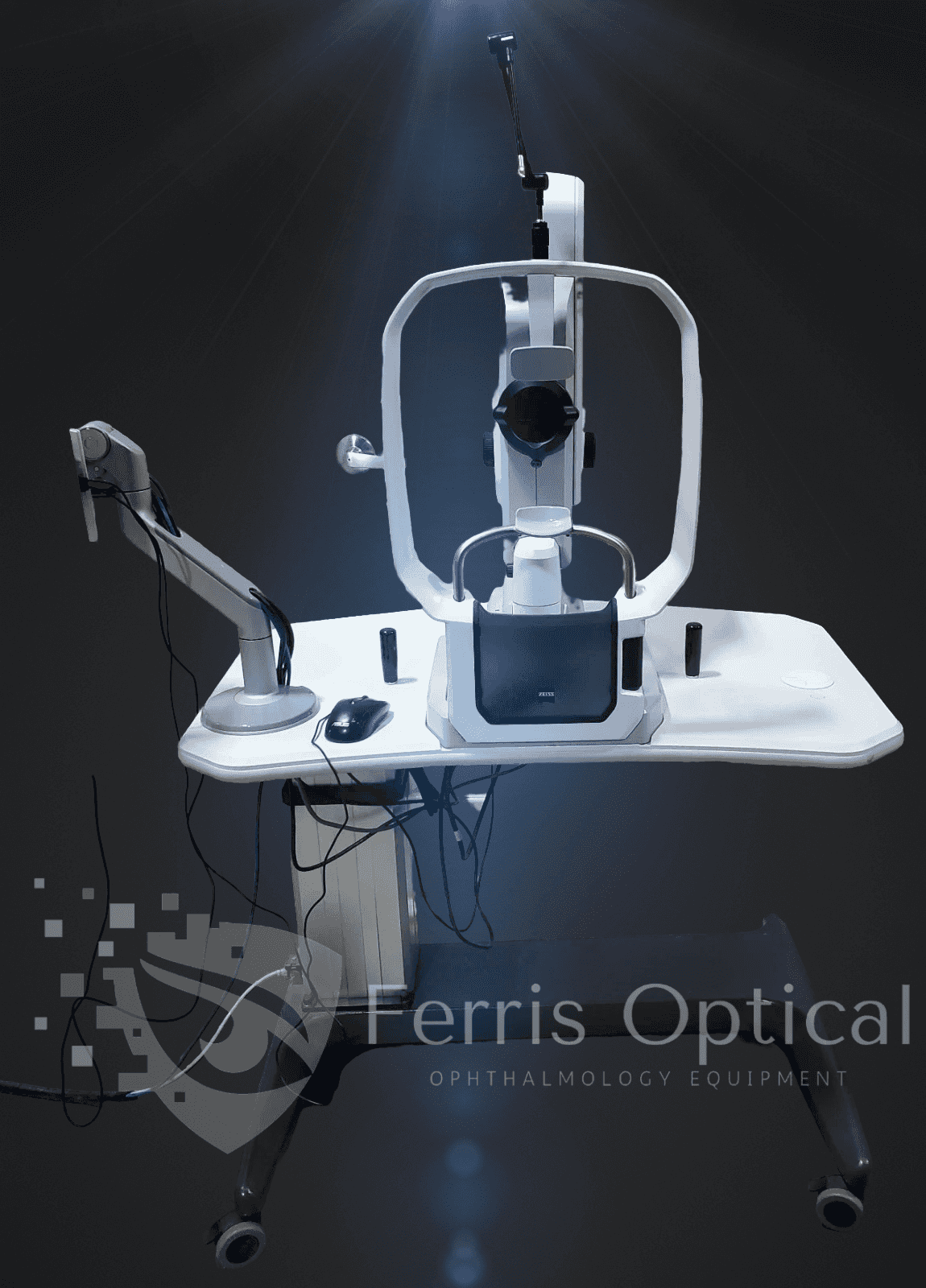
- Zeiss Clarus 500 Ultra Wide Field Retinal Fundus Camera. Fully Tested with Fantastic Unit and Just Removed From Optical Center-Equipment Was Always Serviced By Zeiss.
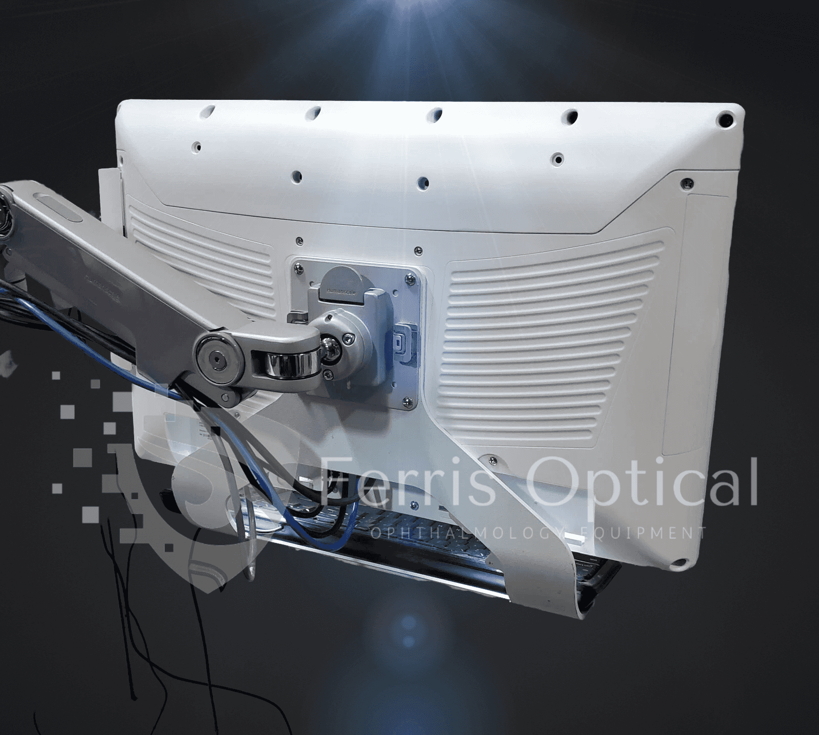
- Onyx Healthcare 22″ All-in-One Computer
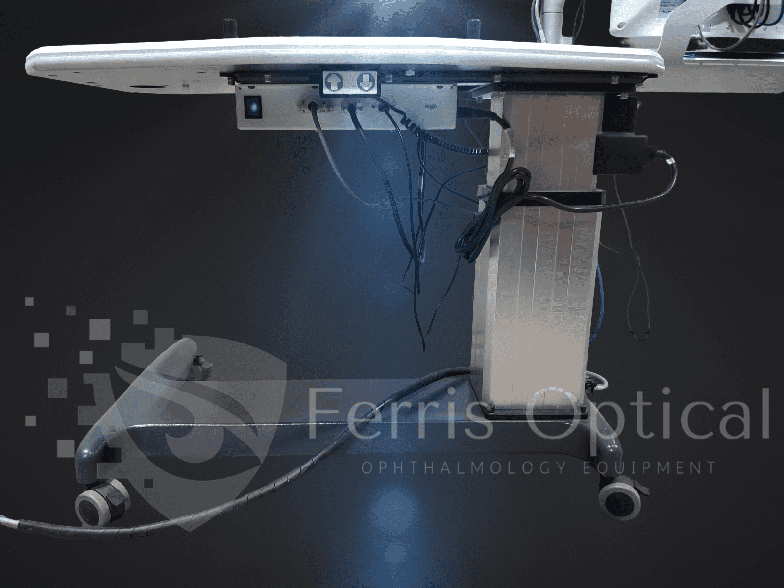
- K2 Tables K2-110C Powered Table
- Microsoft Keyboard and Asus Mouse
- Zeiss Power Table/Manuals/Cover
Software Package:
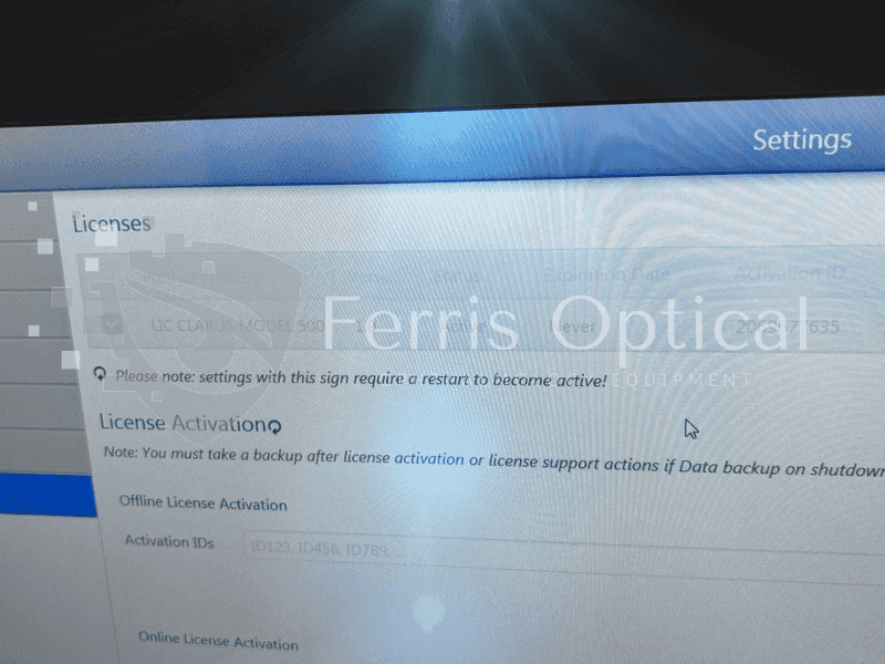
- Firmware Version: 1.2.0.11
- Fundus Camera Version 2.0.9
- FPGA Version 132
PC Specs:
- Processor: Intel(R) Core(TM) i5-6500TE CPU @ 2.30GHz 2.30 GHz
- RAM: 8.00 GB
- Windows 10 Enterprise 64-Bit
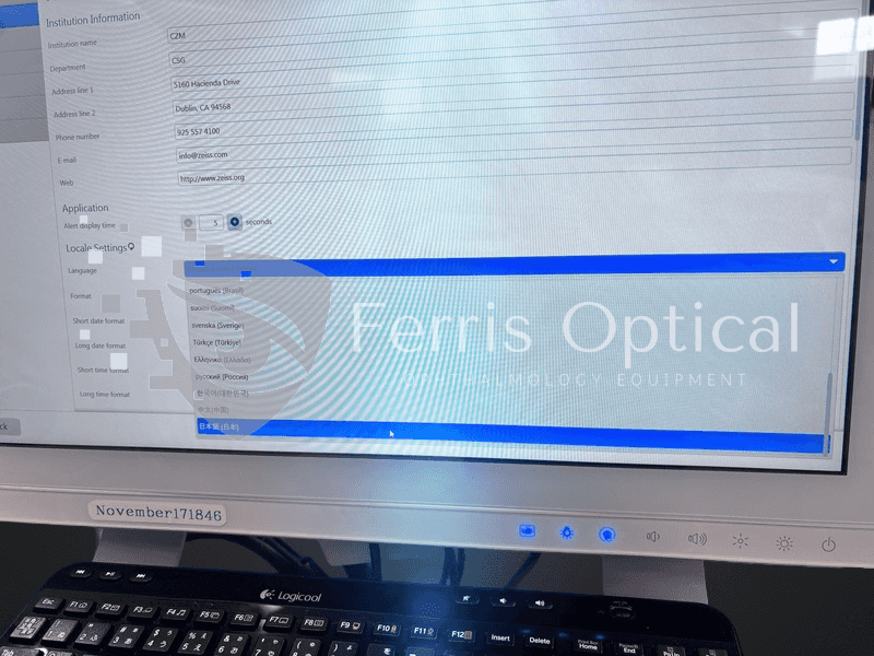
 English
 Spanish
 German
 French