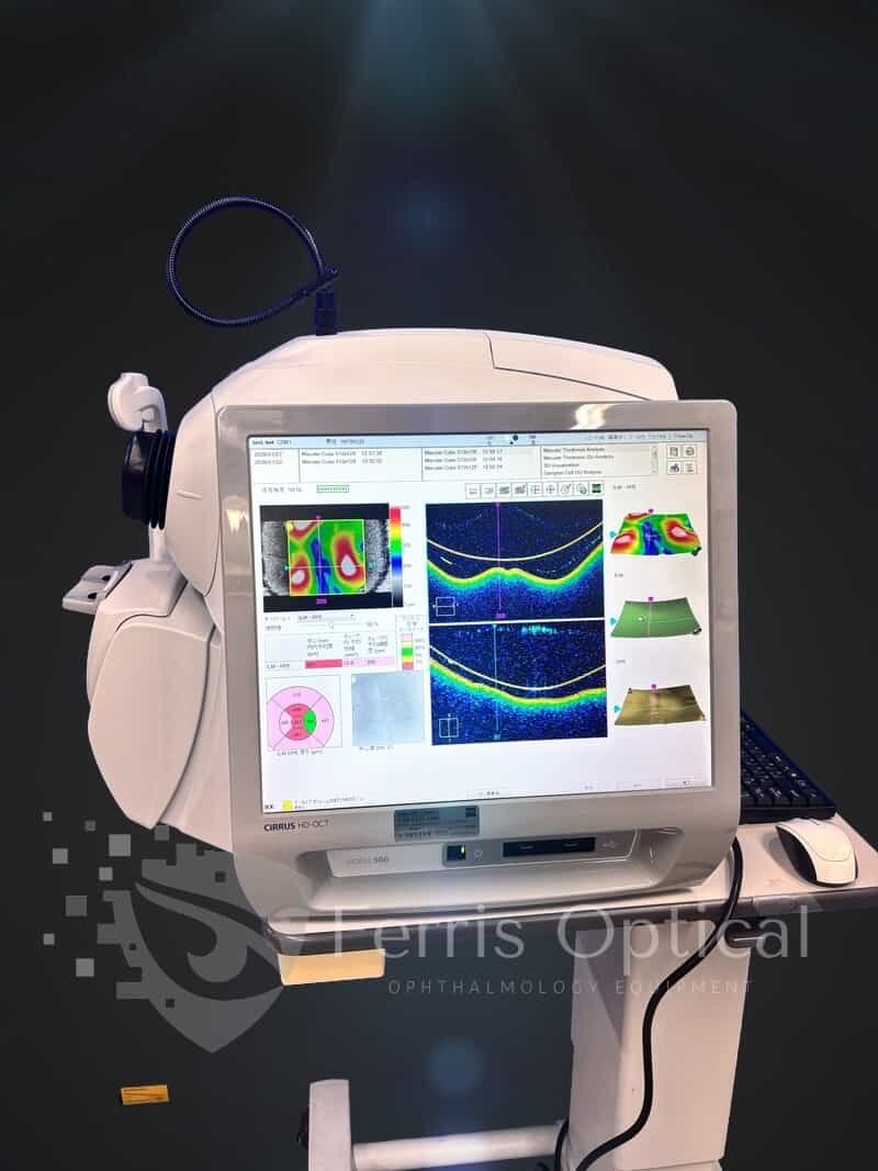
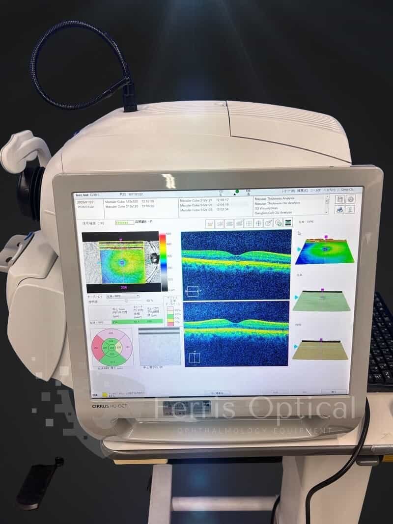
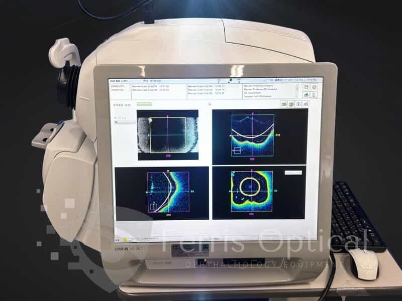
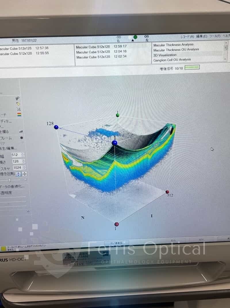
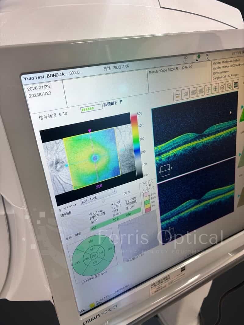
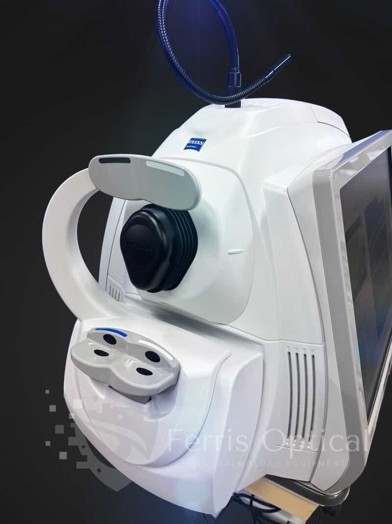
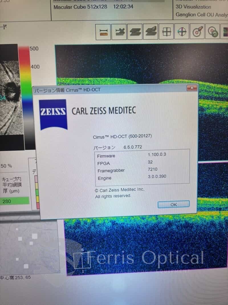
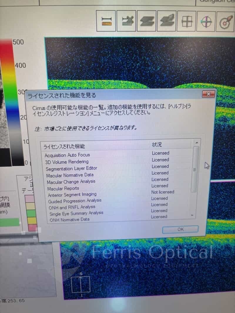
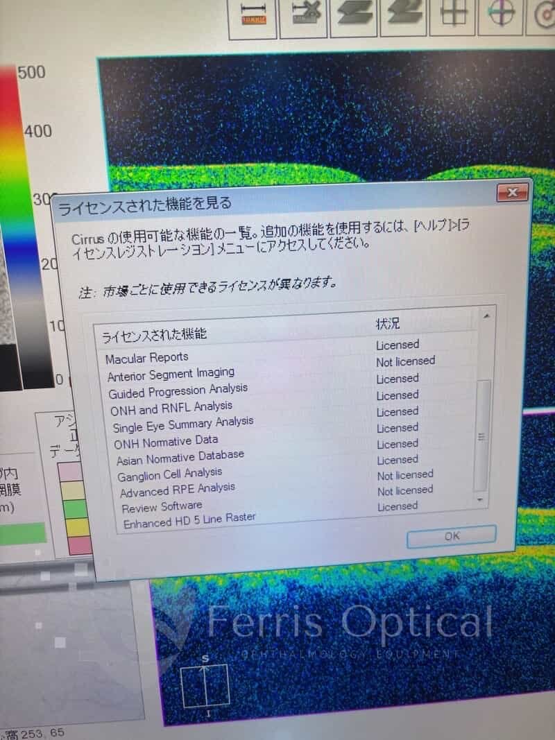
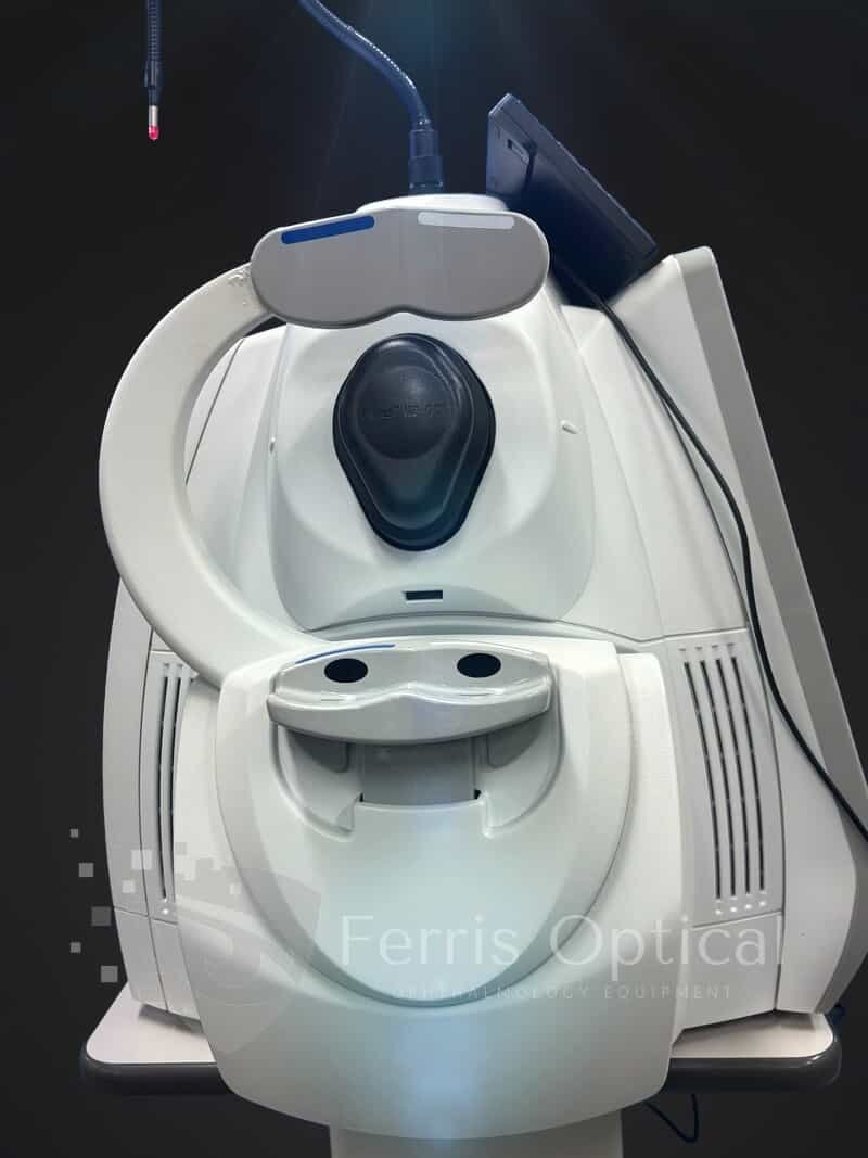
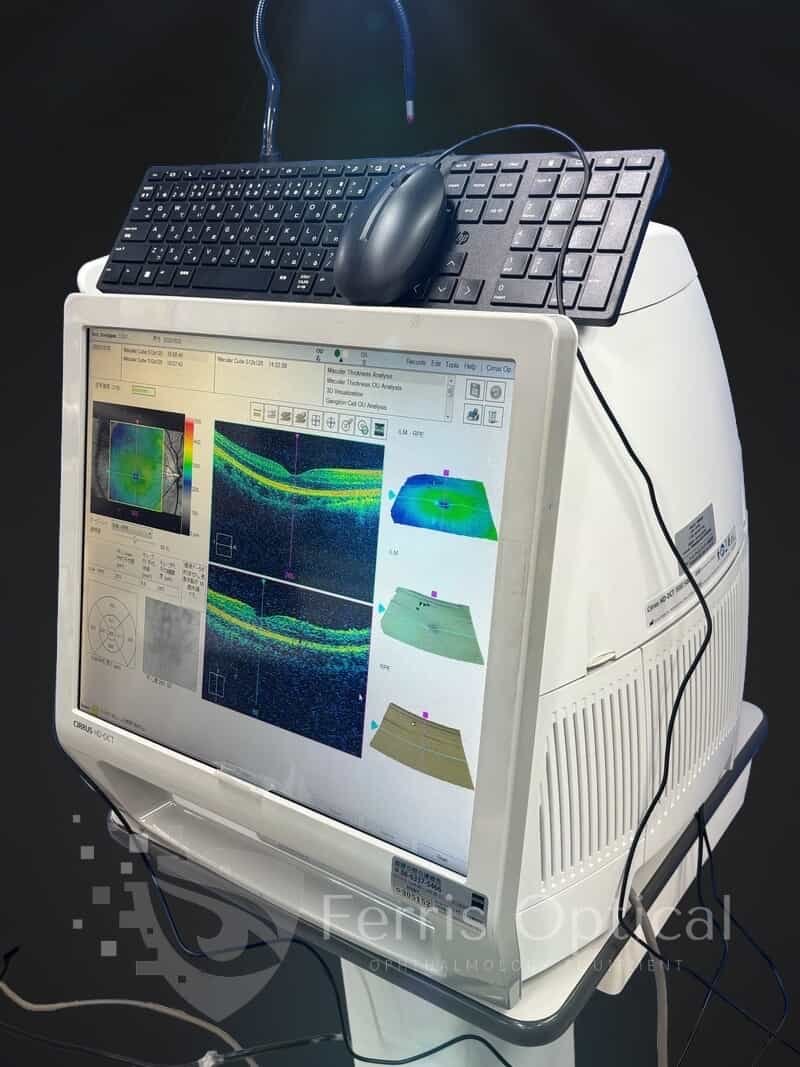
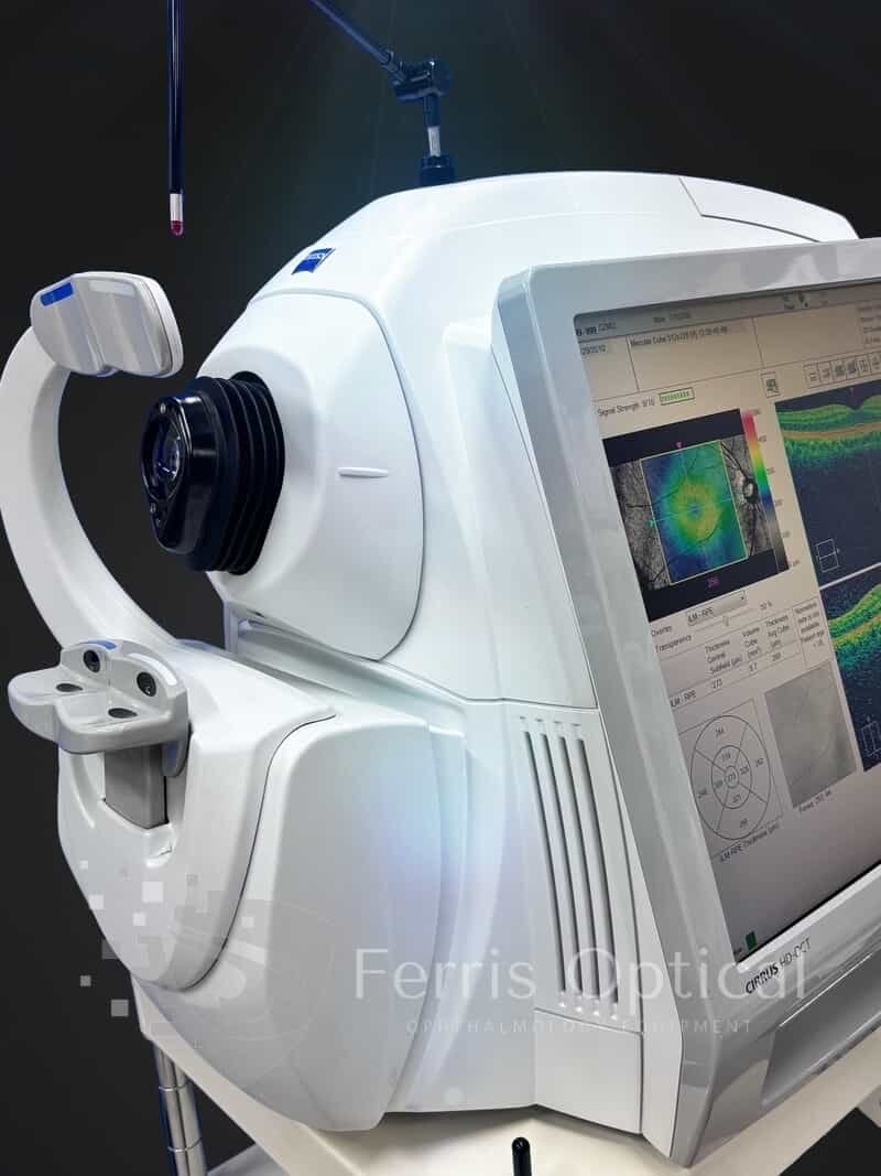
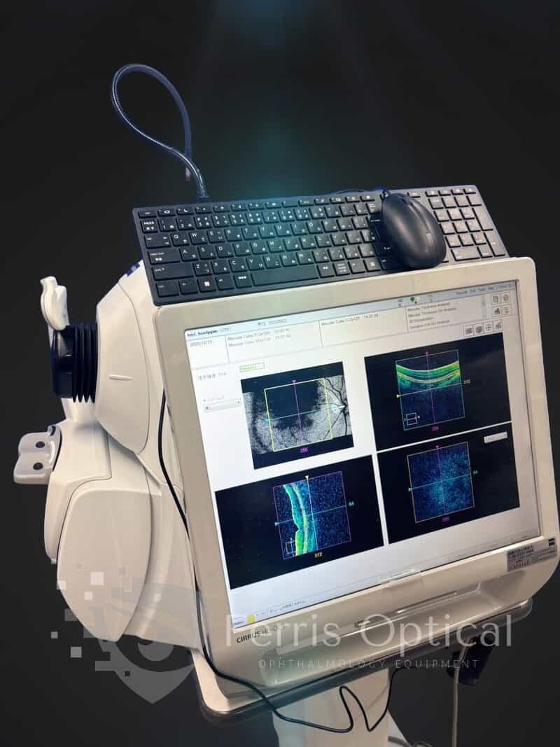
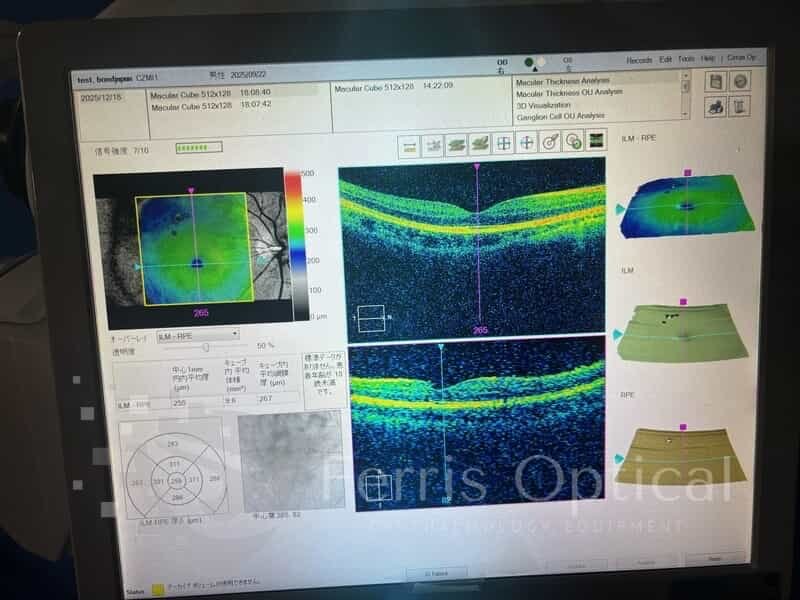
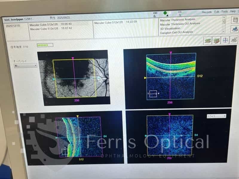
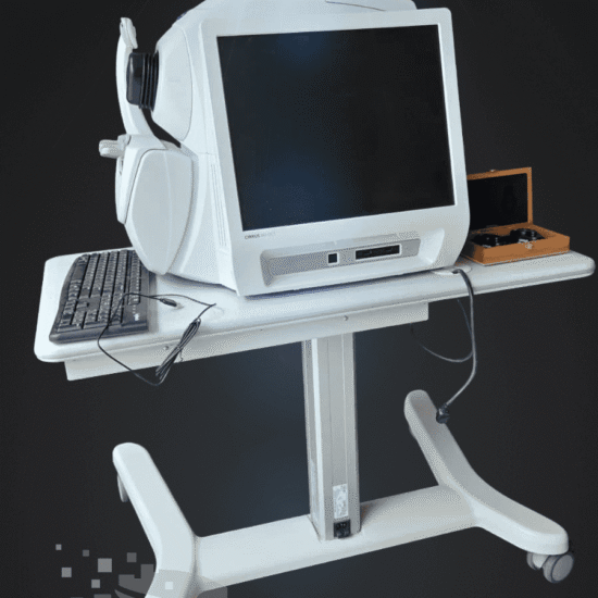
The CIRRUS HD-OCT 500 is purpose-built for optometrists who need essential OCT capabilities and a wide range of clinical applications in a user-friendly platform. This system streamlines the management of glaucoma and retinal disease, supports retina assessment for cataract surgery, and enables anterior segment imaging for corneal disease in modern optometry clinics.
CIRRUS HD-OCT 500 provides optometrists with a practical, easy-to-use solution for comprehensive imaging needs—covering glaucoma and retinal disease management, retinal assessment for cataract surgery, and anterior segment imaging for corneal disease. The new OCT camera delivers rapid fundus image refreshes, improving patient alignment and workflow in busy optometry clinics.
The Zeiss Cirrus HD-OCT 500 is designed for optometrists who want a fast, effective OCT solution for a broad spectrum of retinal and anterior segment diagnostics. The system’s rapid fundus image refresh and intuitive workflow help optimize patient alignment and streamline clinical assessments—improving the quality and efficiency of care in optometry practices.
Features:
- Macular Thickness Normative Data
- Guided Progression Analysis (GPA™)
- Macular Thickness OU Analysis
- Ganglion Cell Analysis
- Macular Thickness and Change Analysis
Zeiss Cirrus 500 OCT — Refurbished Spectral-Domain OCT for Optometrists: GPA and Ganglion Cell Analysis
The Zeiss Cirrus 500 OCT equips optometry clinics with Guided Progression Analysis (GPA), Ganglion Cell Analysis (GCA), and the full suite of Cirrus macular and optic disc protocols.
This clinically proven SD-OCT has been validated in thousands of peer-reviewed glaucoma and retina studies—giving optometrists confidence in normative comparisons and progression statistics at every visit.
The system covers the full spectrum of posterior segment imaging, from RNFL mapping and optic disc cube scans to macular thickness grids and high-definition rasters.
Peer-reviewed research confirms that Guided Progression Analysis (GPA) in the Cirrus 500 OCT enables statistically validated detection of glaucoma progression at any stage—empowering optometrists to deliver evidence-based care and meet co-management documentation standards.
Why Optometry Clinics Choose the Zeiss Cirrus 500 OCT
- Guided Progression Analysis (GPA): Detects statistically significant RNFL thinning between visits and generates clear summary reports—the gold standard for glaucoma progression documentation in optometry clinics.
- Ganglion Cell Analysis (GCA): Maps the macular ganglion cell and inner plexiform layer complex, enabling optometrists to detect early glaucoma changes even before RNFL loss is visible.
- 5-micron axial resolution at 840 nm: Delivers crisp, detailed images for precise RNFL thickness measurement, macular mapping, and AMD monitoring in optometry clinics.
- Full Cirrus protocol suite: Comprehensive macular, optic disc, and anterior segment protocols are built in, supporting all standard clinical workflows for optometry clinics.
- Cirrus normative database: Access age-stratified RNFL and GCC normative values from the most-cited Cirrus reference dataset, supporting confident diagnostics and progression tracking for optometrists.
- Anterior segment capable: An optional anterior segment lens enables optometrists to image the cornea, angle, and anterior chamber without additional equipment.
- Familiar Cirrus interface: Optometrists benefit from the same software platform, protocols, and workflow as other Cirrus models—ensuring a minimal learning curve and easy adoption in any clinic.
Clinical Applications for Optometrists
- Glaucoma progression monitoring: GPA identifies statistically significant RNFL changes between visits, supported by the Cirrus normative database—helping optometrists track disease progression and guide management decisions.
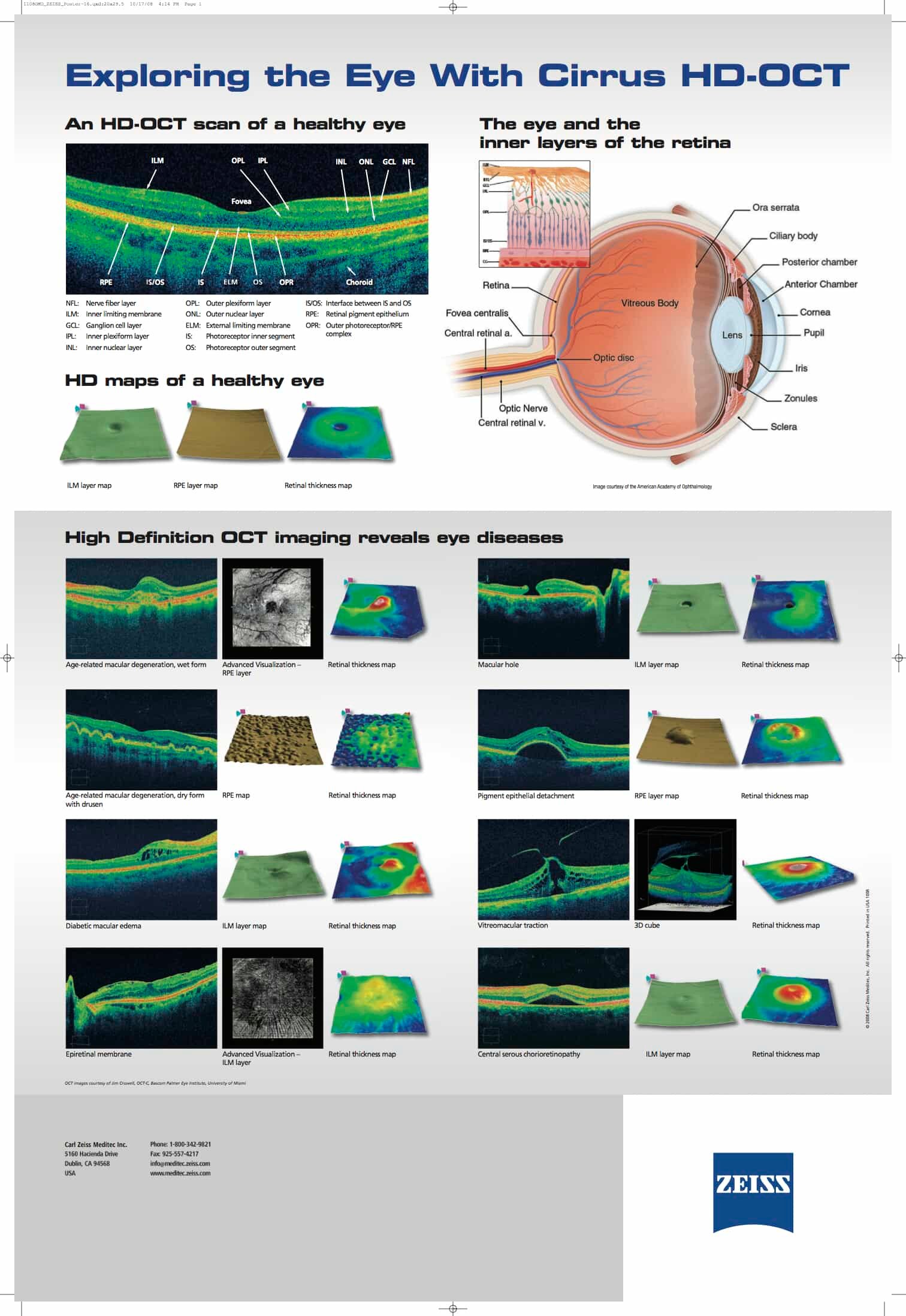
- Macular disease surveillance: Monitor AMD, DME, epiretinal membranes, and vascular occlusions using detailed macular imaging and thickness maps to support long-term patient care in optometry clinics.
- Optic disc documentation: Capture the full disc and peripapillary RNFL in a single scan for baseline and follow-up documentation—supporting comprehensive care in optometry clinics.
- Pre- and post-operative evaluation: Use macular thickness maps and RNFL status to support cataract, vitreoretinal, and glaucoma surgical planning, co-management, and outcomes tracking in optometry clinics.
- Anterior segment imaging: Perform corneal and anterior chamber assessments with the optional lens to expand diagnostic capabilities in optometry clinics.
Who Benefits in Optometry
- Optometrists specializing in glaucoma: GPA and GCA deliver the data and documentation needed for rigorous progression monitoring and managed care reporting.
- Comprehensive optometry practices: Cover RNFL analysis, macular screening, and optic disc documentation for your full patient panel with a single instrument.
- Optometry clinics upgrading from older OCT platforms: Benefit from the Cirrus normative database and GPA tools for a significant clinical advancement over first-generation instruments.
- Optometrists entering glaucoma co-management: Cirrus is accepted by major co-management reporting frameworks, and GPA provides the documentation needed for collaborative care.
- Multi-location optometry groups: Standardize your platform, protocols, and normative values across all sites with the Cirrus 500 for seamless care and consistent diagnostics.
Specifications:
OCT Imaging
- Methodology: Spectral Domain OCT
- Optical source: Superluminescent diode (SLD), 840 nm
- Scan Speed: 27k-68K A-scans per second
- A-scan: mm (in tissue), 1204
- Axial resolution: 5 m (in tissue)
- Transverse resolution: 15 m (in tissue)
Fundus Imaging
- Methodology: Line scanning ophthalmoscope (LSO)
- Live fundus image: During alignment and during OCT scan
- Optical source: Superluminescent diode (SLD), 750 nm
- Field of view: 36 degrees W x 30 degrees H
- Frame rate: > 20 Hz
- Transverse resolution: 25 m (in tissue)
Iris Imaging
- Methodology: CCD camera
- Resolution: 1280 x 1024
- Live iris image: During alignment
Includes
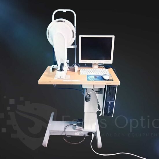
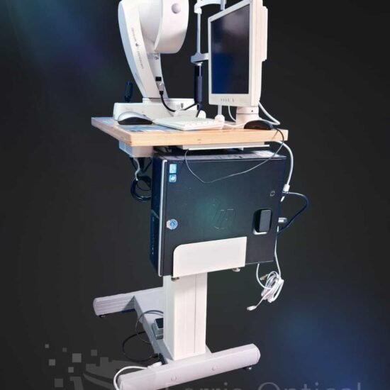
- Carl Zeiss Cirrus 500 HD-OCT Main Unit
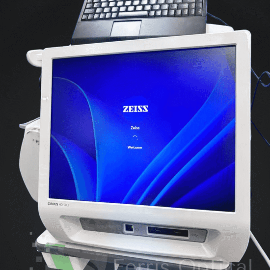
- Integrated Windows 10 PC (Pre-installed software)
- Power table
- Dust cover
- Power cable
- 6 Month warranty
- Keyboard with touch cursor
- Mouse
Reference
 English
 Spanish
 German
 French